Squamous cell carcinoma L premaxillary with perineural tumor
Findings:
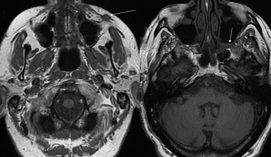
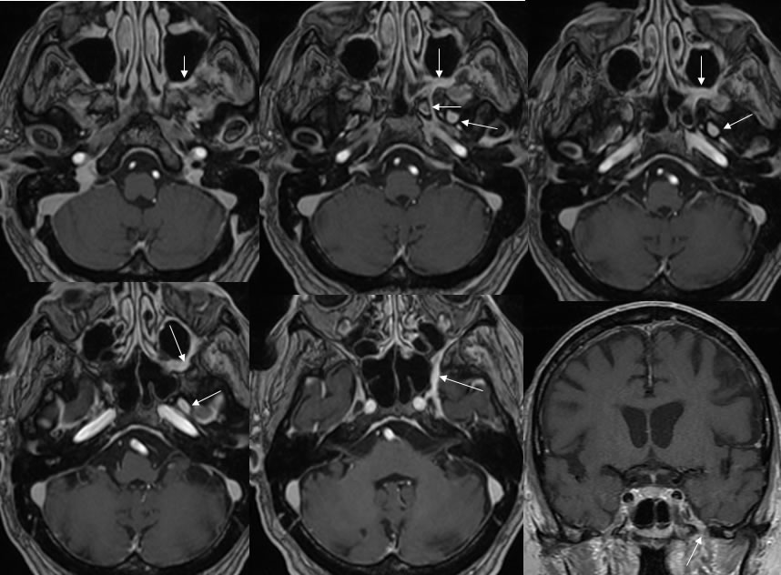
A poorly defined spiculated mass in the left premaxillary region is demarcated by the arrow on the T1 precontrast image. The second T1 precontrast image arrow demarcates abnormal signal within the pterygomaxillary fissure with loss of the normal fat signal. Additional arrows on the post contrast images demarcate zones of abnormal enhancement within the pterygomaxillary fissure, vidian canal, foramen rotundum, and foramen ovale, which is also seen on the coronal postcontrast image through foramen ovale.
Discussion/Differential Diagnosis:
BACK TO
MAIN PAGE