Recurrent Adenoid Cystic Carcinoma with Perineural Tumor
Findings:
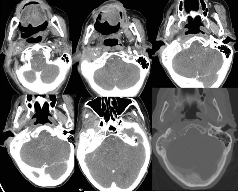
Multiple axial neck CT images with contrast demonstrate abnormal infiltrative soft tissue opacity adjacent to the right mastoid region extending into the masticator space and encasing multiple vascular structures. There is abnormal opacification of the right mastoid air cells and infiltrative soft tissue opacity in the right retroauricular region. The bone window image demonstrates irregular demineralization of the right aspect of the clivus and mastoid air cells with zones of bone destruction. Intracranial heterogeneity is poorly characterized on the CT images, with apparent mass affect on the fourth ventricle. Abnormal vascularity projects over the lower brainstem and cerebellum.
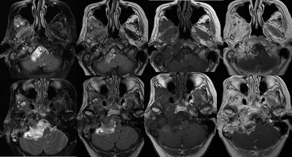
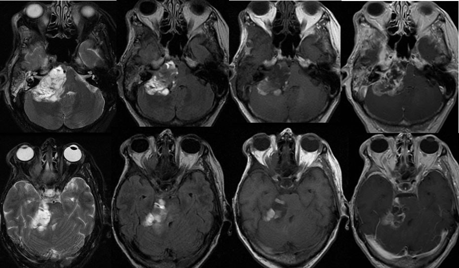
The MRI images demonstrate an extensive poorly defined heterogeneously enhancing complex cystic and solid mass within the right posterior fossa which appears extraaxial, causing mass effect on the brainstem and fourth ventricular effacement. The margins of the mass are infiltrative adjacent to brain parenchyma. Extensive abnormal enhancement encases the right internal carotid and infiltrates the right masticator space. Abnormal marrow signal involves the right mastoid region and the right aspect of the clivus. The mass has very complex signal characteristics with likely some hemorrhagic changes intracranially. Additional homogeneously enhancing mass component extends laterally from the right mastoid air cells. In some regions, tumor and inflammatory disease are difficult to separate within the mastoid air cells. Postoperative changes of partial right parotidectomy are present.
Discussion/Differential Diagnosis:
BACK TO
MAIN PAGE