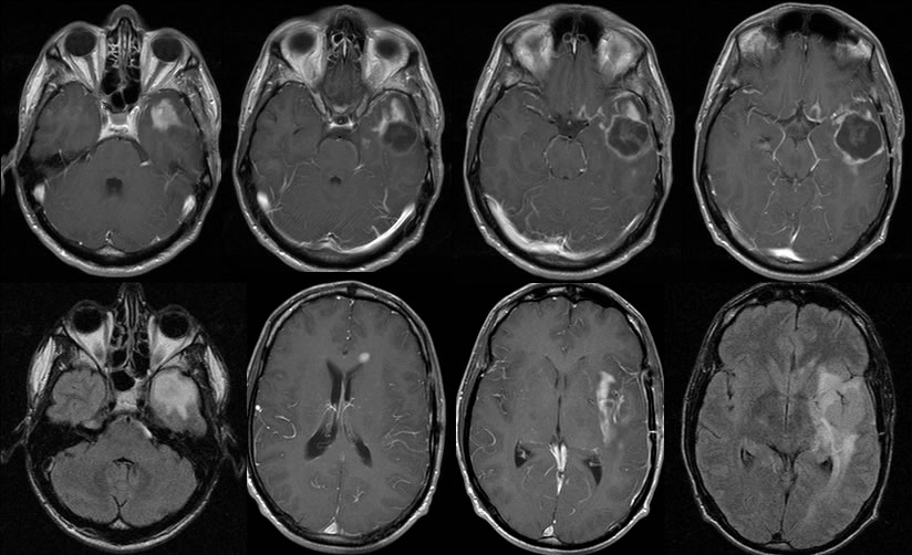
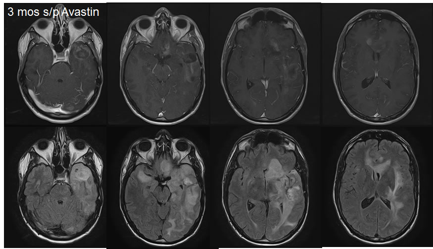
Recurrent GBM with subarachnoid and subependymal dissemination, pseudoresponse after Avastin.
Findings:
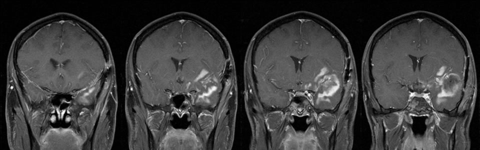
Multiple MRI images demonstrate postoperative changes of left temporal craniotomy. An irregular surgical defect is present in the left temporal lobe with peripheral irregular enhancement. Poorly defined nodular FLAIR signal alteration extends along the margins of the surgical defect into the anterior left temporal lobe. The nodular zones of poorly defined enhancement extend into the left insula and left frontal periventricular region. There is also extensive abnormal leptomeningeal enhancement along the left temporal and left inferior frontal lobes.
Follow up images after Avastin therapy demonstrate overall decreased enhancement, but the enhancement is more extensive and more poorly defined. The nodular FLAIR signal alterations are substantially progressive with evolving effacement of the left lateral ventricle. The anterior corpus callosum is now expanded with extensive nodular signal alteration and poorly defined enhancement.
Discussion/Differential Diagnosis:
BACK TO
MAIN PAGE