CNS lymphoma with perivascular disease
Findings:
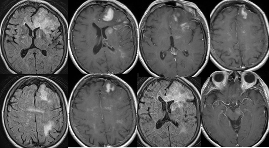
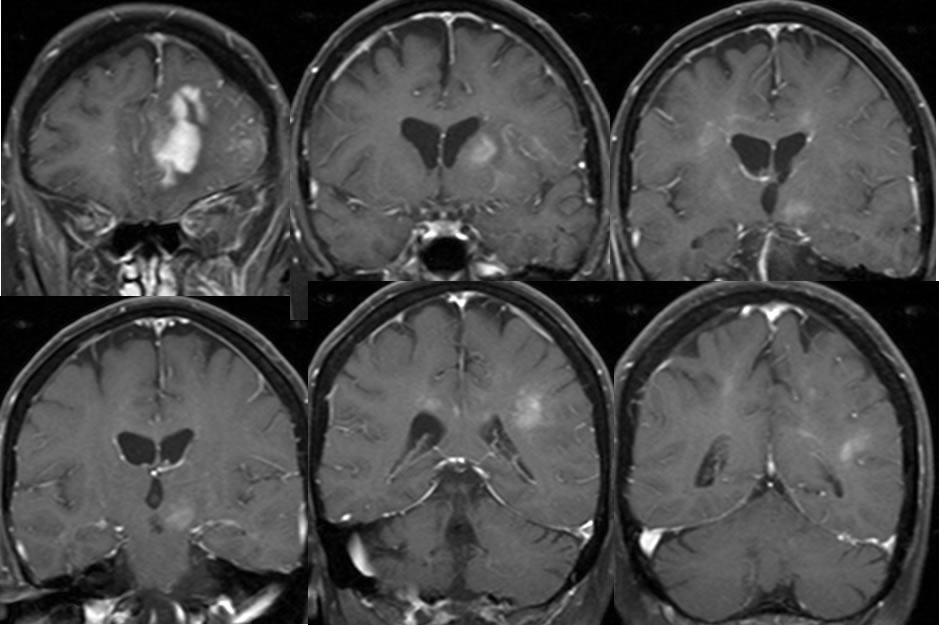
Multiple MRI images demonstrate a confluent poorly defined enhancing mass with near homogeneous enhancement in the left frontal lobe, with extensive surrounding FLAIR signal alteration and mild mass effect . Additional poorly defined linear foci of abnormal enhancement and FLAIR signal alteration extend along perivascular spaces in the bilateral centrum semiovale, with bandlike linear enhancement crossing the corpus callosum. Additional poorly defined enhancement extends into the left cerebral peduncle and left caudate nucleus. There is very little mass effect overall for the extent of disease.
Discussion/Differential Diagnosis:
BACK TO
MAIN PAGE