CNS lymphoma
Findings:
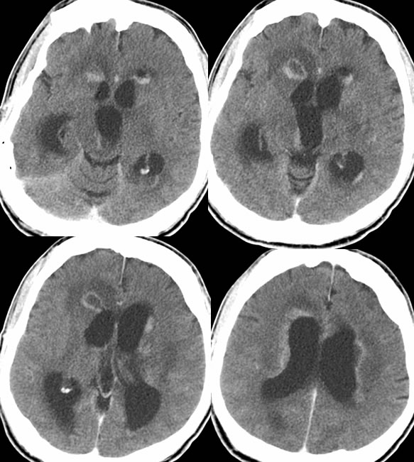
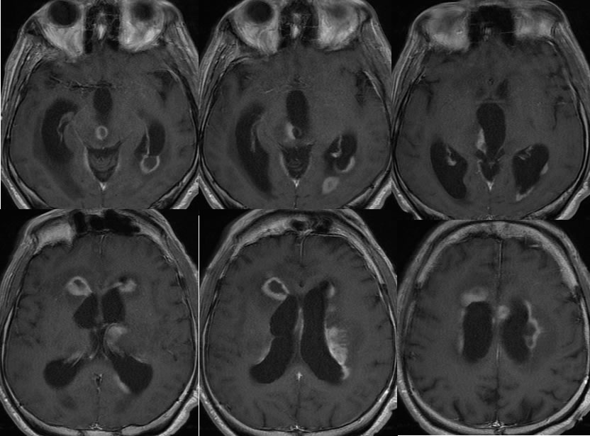
Contrast enhanced CT and MRI images demonstrate dilated ventricles due to hydrocephalus. There is extensive poorly defined subependymal nodular enhancement along the margins of the bilateral lateral ventricles and posterior margin of the third ventricle. Periventricular CT low attenuation represents a combination vasogenic edema and transependymal CSF flow.
Discussion/Differential Diagnosis:
BACK TO
MAIN PAGE