Metastatic Melanoma, leptomeningeal
Findings:
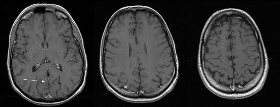
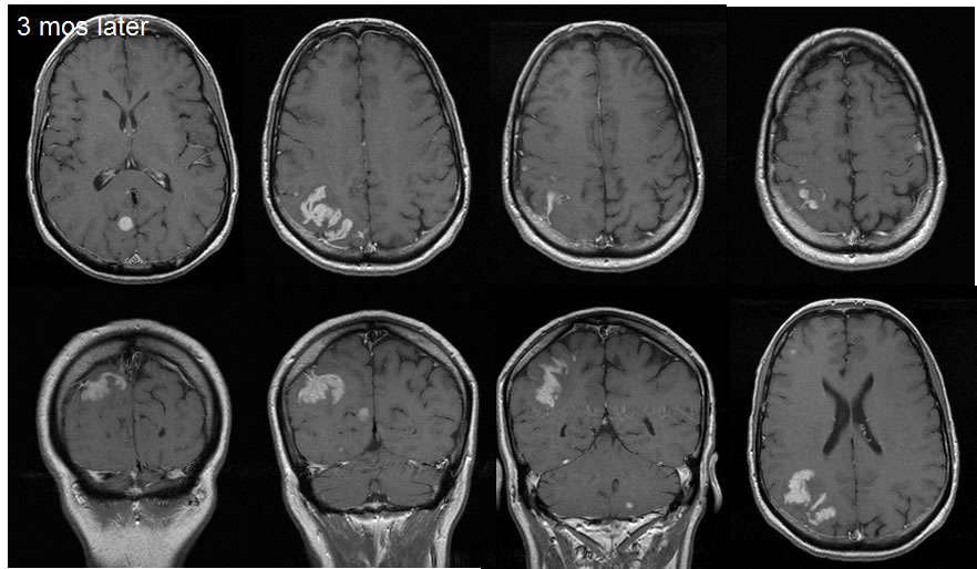
The initial MRI T1 post contrast images demonstrate small enhancing lesions within the left frontal cortex, right parietal cortex, and right parietooccipital region. An arrow demarcates the subtle right parietooccipital lesion. The follow up images three months later demonstrate development of extensive leptomeningeal enhancement in the right parietal lobe. Other enhancing lesions have increased in size. No significant mass effect is demonstrated.
Discussion/Differential Diagnosis:
BACK TO
MAIN PAGE