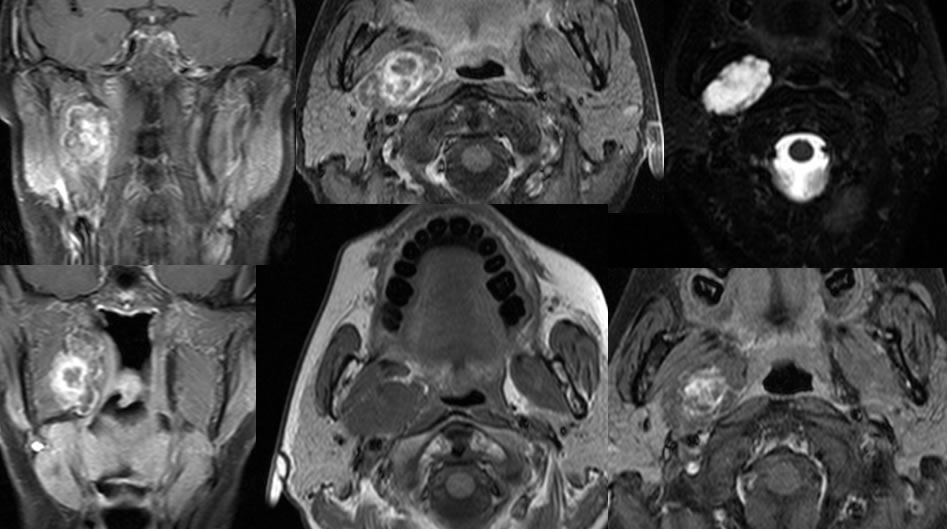
Benign Mixed Tumor, Deep Lobe R Parotid
Findings:
T1 post contrast images with fat saturation demonstrate a multilobulated heterogeneously enhancing mass within the deep lobe right parotid gland. The lesion demonstrates overall marked T2 hyperintensity with slight heterogeneity. The central zone of the mass demonstrates intense peripheral enhancement with a small zone of cystic change or central necrosis. The mass is nearly isointense to muscle on T1 pre-contrast imaging.
Discussion/Differential Diagnosis:
BACK TO
MAIN PAGE