Subarachnoid Metastases, Melanoma
Findings:
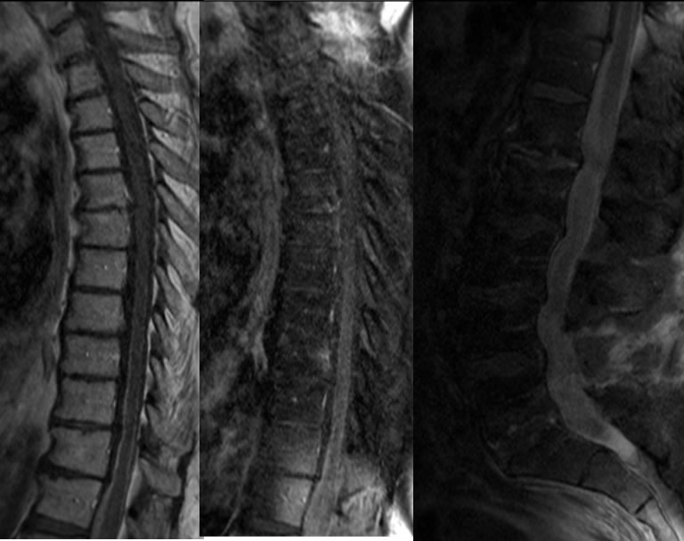
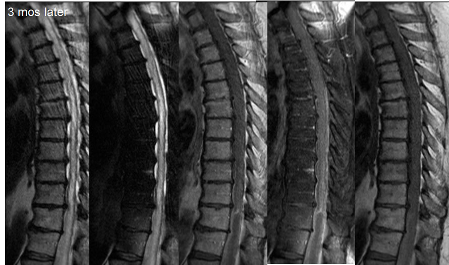
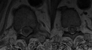
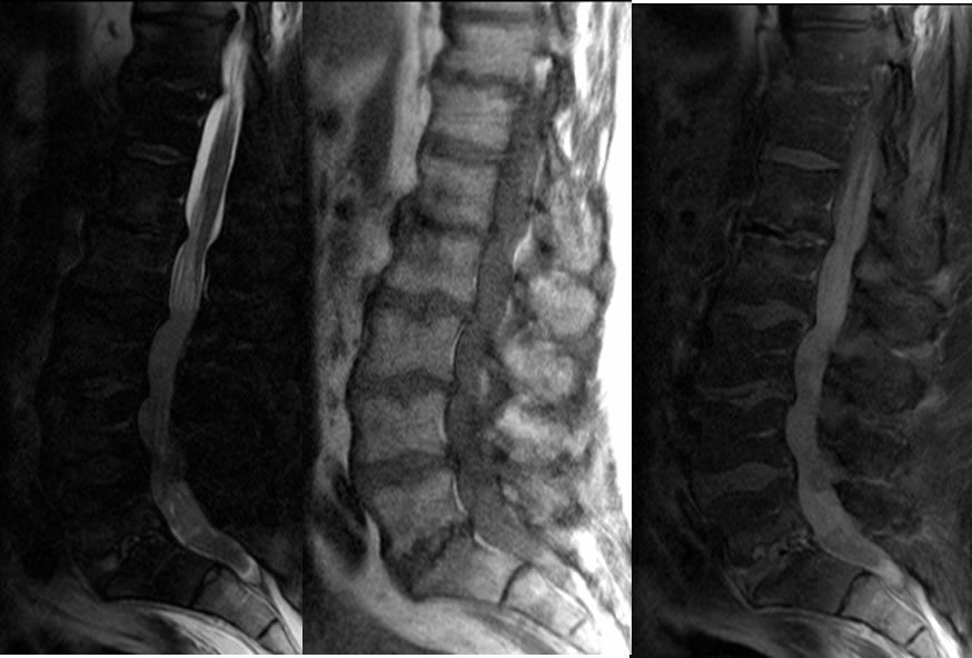
Sagittal T1 weighted images without and with fat saturation through the thoracic spine demonstrate linear nodular enhancement coating the distal thoracic cord and conus. Sagittal imaging of the lumbar spine with fat saturation and contrast demonstrates extensive abnormal leptomeningeal enhancement filling the thecal sac. The follow up images three months later demonstrate progression of disease with development of extensive cord edema signal. Superimposed advanced multilevel degenerative disc disease is present.
Discussion/Differential Diagnosis:
BACK TO
MAIN PAGE