Hemangioblastoma
Findings:
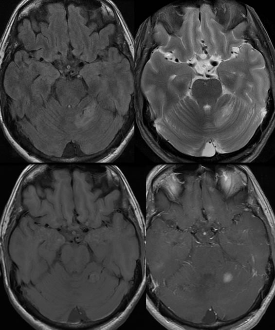
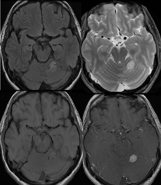
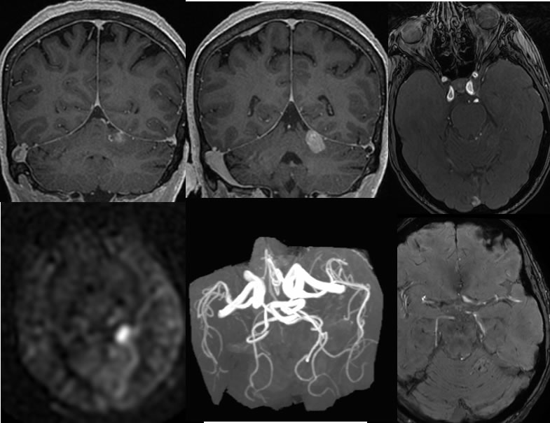
Multiple MRI images demonstrate a nodular soft tissue mass within the superior right cerebellum with linear foci of decreased signal due to flow voids. Mild surrounding vasogenic edema is present. No significant arterial flow signal is seen in this region on the MRA. Hyperperfusion is seen in this region on arterial spin labeling. No significant hemorrhagic staining is visible.
Discussion/Differential Diagnosis:
BACK TO
MAIN PAGE