Chiari 3 malformation, status post remote repair
Findings:
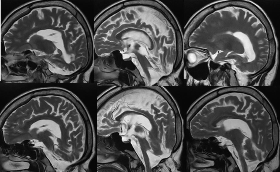
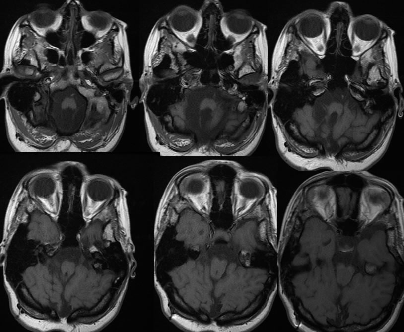
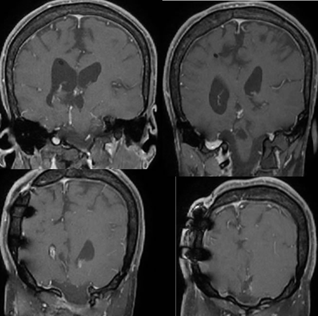
Sagittal T2 weighted MRI images demonstrate no visible cerebellum with brainstem atrophy and distortion of the tectum. The axial T-1 MRI images demonstrate a wide well-defined defect in the midline occipital bones with flaring at the margins indicating a congenital dysgraphia. The occipital lobes are displaced inferiorly into the posterior fossa. A well-defined fourth ventricle is not visible. Susceptibility artifact from additional postoperative changes along the right parietal calvarium are present. A shunt catheter is incompletely included. Superimposed skull base fusion anomalies are present incompletely included, including atlantooccipital fusion.
Discussion/Differential Diagnosis:
BACK TO
MAIN PAGE