Cavernoma and Developmental Venous Anomaly
Findings:
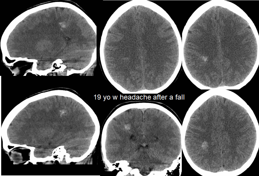
Multiple CT images demonstrate a multi lobulated irregular hyperdense structure within the deep right parietal white matter. With the history of trauma, please note that there is no surrounding vasogenic edema or mass effect. While the hyperdense structure has Hounsfield units in the region of hemorrhage, the location and configuration would be very unusual for a zone of acute parenchymal hemorrhage.
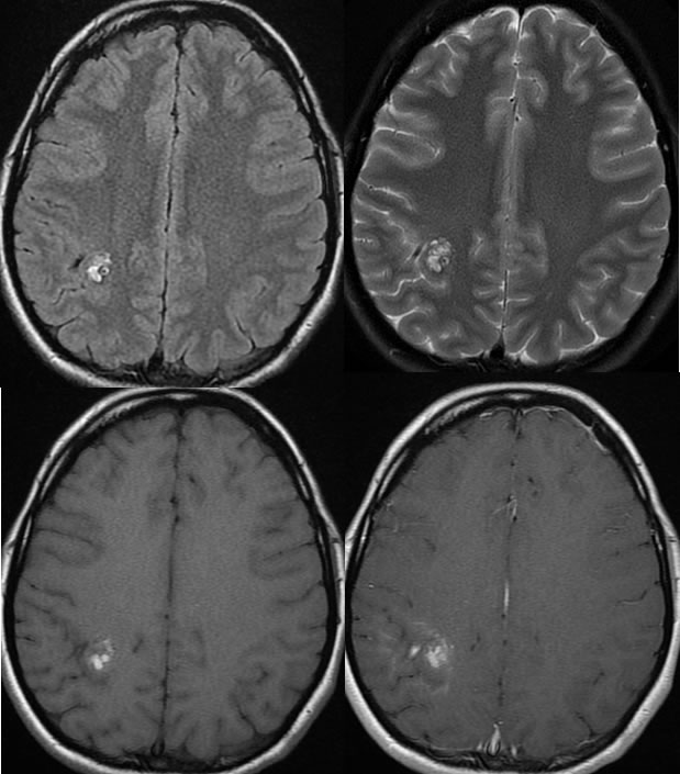
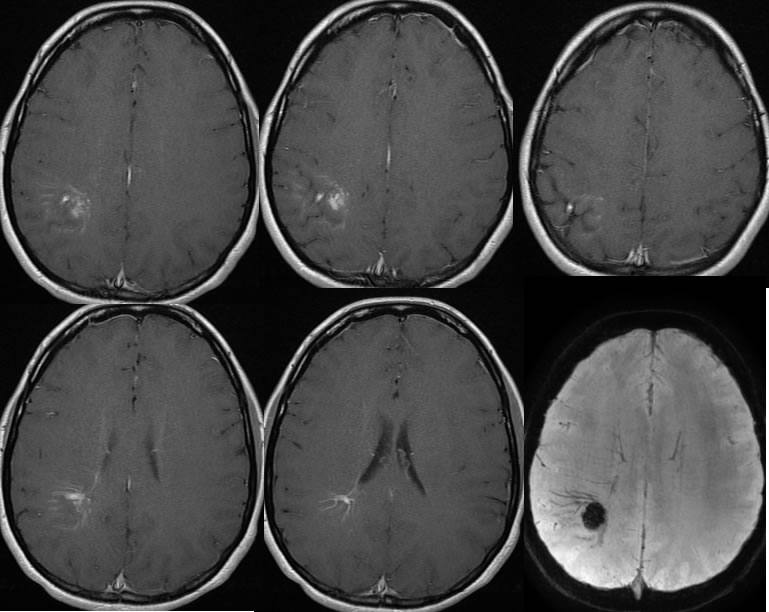
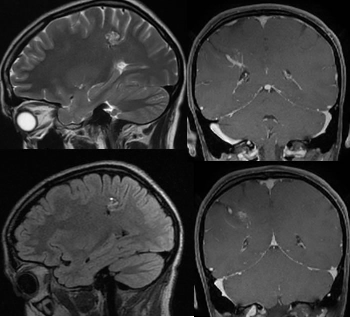
MRI images demonstrate a multilobulated signal alteration in the same region which has zones of T1 precontrast hyperintensity and multiple septations. Dense susceptibility artifact is visible in this region on the susceptibility weighted sequence. The lesion as overall T2 hyperintense with a relatively hypointense irregular rim. Stippled internal T2 hyperintensities are present. There is a superimposed large venous structure in the region of the hemorrhagic lesion which has branching tributaries into a larger trunk, indicating a large developmental venous anomaly, which commonly coexists with a cavernoma.
MRI images demonstrate a multilobulated signal alteration in the same region which has zones of T1 precontrast hyperintensity and multiple septations. Dense susceptibility artifact is visible in this region on the susceptibility weighted sequence. The lesion as overall T2 hyperintense with a relatively hypointense irregular rim. Stippled internal T2 hyperintensities are present. There is a superimposed large venous structure in the region of the hemorrhagic lesion which has branching tributaries into a larger trunk, indicating a large developmental venous anomaly, which commonly coexists with a cavernoma.
Discussion/Differential Diagnosis:
BACK TO
MAIN PAGE