Secondary CNS Lymphoma
Findings:
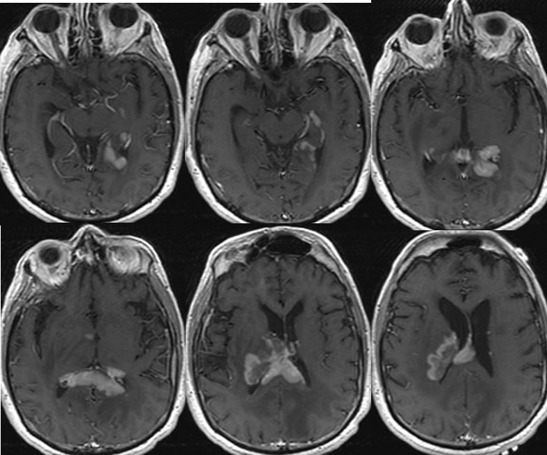
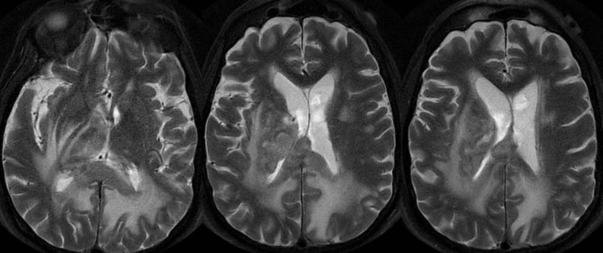
Multiple T1 post contrast images demonstrate irregular nodular near homogeneously enhancing masses infiltrating the corpus callosum splenium, right corona radiata, left medial temporal lobe, and right medial thalamus. There is extensive surrounding T1 hypointensity due to vasogenic edema. The multiple masses are relatively hypointense on T2 weighted imaging indicating cellularity.
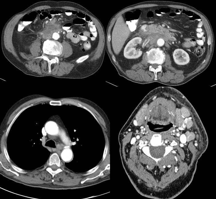
CT imaging of the abdomen, chest, and neck demonstrates multifocal adenopathy with a confluent poorly defined nodular mass in the retroperitoneum encasing the aorta and other structures.
Discussion/Differential Diagnosis:
BACK TO
MAIN PAGE