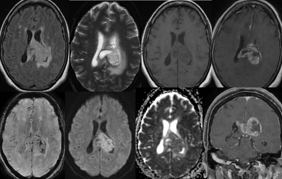
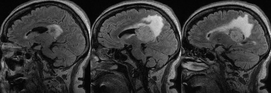
Glioblastoma Multiforme
Findings:
Multiple MRI images demonstrate a poorly defined multilobulated peripherally enhancing mass involving the corpus callosum posterior body asymmetric on the left. The mass crosses midline. There is moderate surrounding signal alteration due to vasogenic edema or nonenhancing tumor. The mass shows spotty zones of hemorrhagic staining on the susceptibility weighted sequence. Restricted diffusion is seen which also involves peripheral aspects of the mass. Some of the centrally necrotic zones do not demonstrate restricted diffusion. The tumor has an irregular mostly solid configuration on the sagittal FLAIR sequence. There is very little mass effect for the size of the lesion.
Discussion/Differential Diagnosis:
BACK TO
MAIN PAGE