Recurrent GBM, perivascular
Findings:
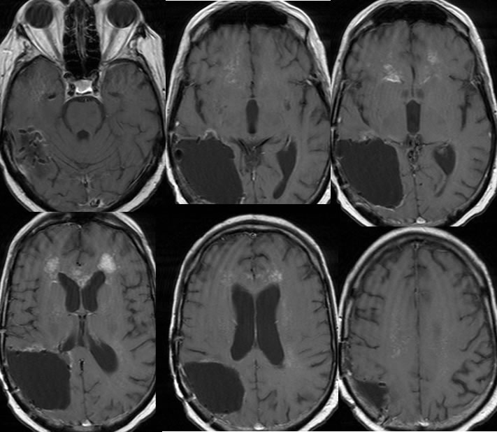
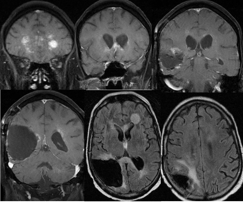
Axial T1 post contrast, coronal T1 post contrast with fat saturation, and axial FLAIR images demonstrate postoperative changes of right parietal craniotomy with a large surgical defect lined by radiation seeds in the right parietal lobe. Spotty irregular zones of abnormal enhancement are present along the margins of the surgical cavity which are most prominent superiorly. Superimposed more extensive linear zones of abnormal enhancement are present in the bilateral frontal white matter and anterior corpus callosum, with some confluent zones of enhancement in the frontal periventricular regions left greater than right. The radiating pattern of abnormal enhancement also extends into The bilateral centrum semiovale along perivascular spaces. There is nodular FLAIR hyperintensity in the region of the largest zones of enhancement. Very little FLAIR hyperintensity is present associated with many of the linear zones.
Discussion/Differential Diagnosis:
BACK TO
MAIN PAGE