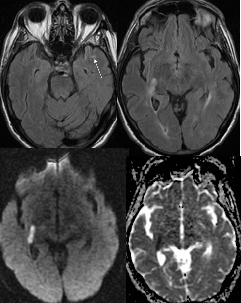
Multicentric GBM, rapidly progressive
Findings:
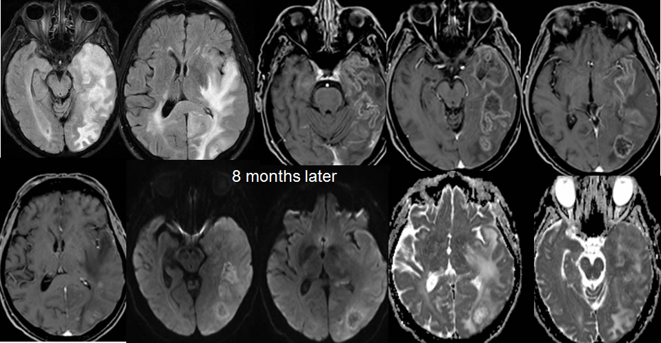
Axial FLAIR and diffusion images demonstrate a nodular zone of diffusion restriction in the posterior right subinsular region. There is a subtle zone of FLAIR hyperintensity in the anterior left temporal lobe, demarcated by the arrow. The follow up images eight months later demonstrate development of multifocal poorly defined nodular zones of abnormal enhancement throughout the left temporal and parietal lobes associated with peripheral diffusion restriction , Mild to moderate mass effect, and extensive surrounding FLAIR signal alteration. Many of these lesions involve overlying cortex with a zone of gyral swelling in the left parietal parasagittal region.
Discussion/Differential Diagnosis:
BACK TO
MAIN PAGE