
Orbital Lymphoma, Secondary CNS Lymphoma
Findings:
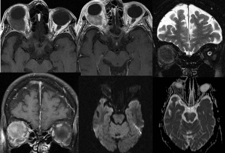
Multiple MRI images of the orbits demonstrate a heterogeneously enhancing mass within the intraconal right orbit which is inseparable from the posterior margin of the right globe and the extraocular muscles. The mass demonstrates decreased T2 signal due to a cellular lesion with mild peripheral diffusion hyperintensity. MRV and MRA images demonstrate no abnormal vascularity in the region of the mass.
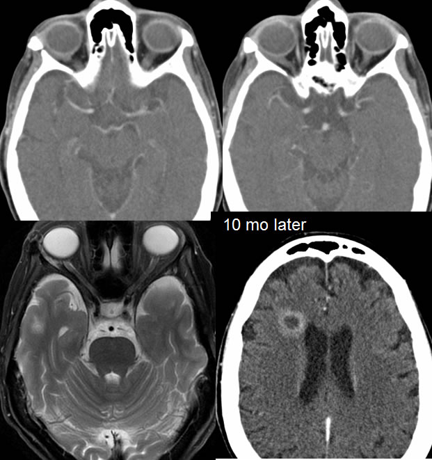
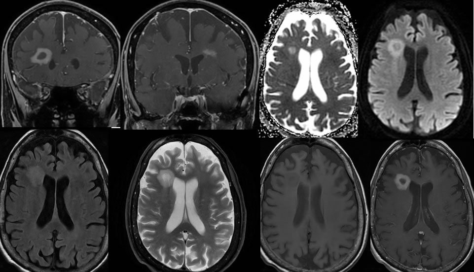
10 months later, CT images demonstrate near complete resolution of the right orbital mass with a mild amount of linear T2 hyperintensity in the region. A poorly defined peripherally enhancing mass with peripheral intermediate restricted diffusion has developed within the deep right frontal white matter with no significant mass effect or surrounding edema. An additional bandlike enhancing focus has developed in the left centrum semiovale.
Discussion/Differential Diagnosis:
BACK TO
MAIN PAGE