CNS Lymphoma
Findings:
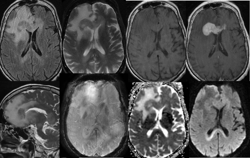
Multiple MRI images demonstrate an irregular solid enhancing mass involving the right frontal white matter and crossing the anterior corpus callosum. This mass demonstrates T2 hypointensity and intermediate restricted diffusion. Moderate surrounding vasogenic edema and mild localized mass effect is present with partial effacement of the right frontal horn. Additional patchy signal abnormalities involve the corpus callosum splenuim with mild expansion and diffusion restriction in the region, but no abnormal enhancement.
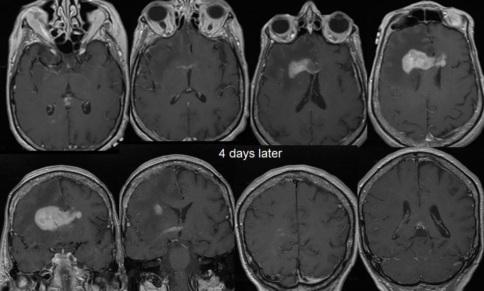
Four days later, the dominant mass has a slightly greater extent into the left frontal white matter. Multiple additional poorly defined zones of abnormal enhancement have developed including along the anterior commissure and in the deep right parietal white matter.
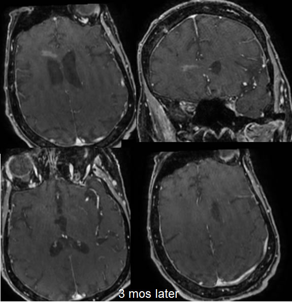
Images performed three months later demonstrate a dramatic improvement of the abnormal enhancement, with mild residual bandlike enhancement in the anterior corpus callosum.
Discussion/Differential Diagnosis:
BACK TO
MAIN PAGE