Spinal Cord Infarction
Findings:
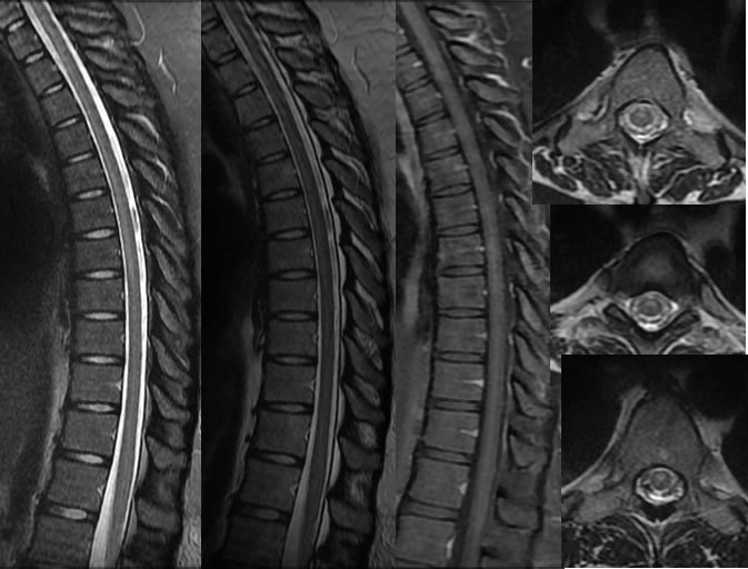
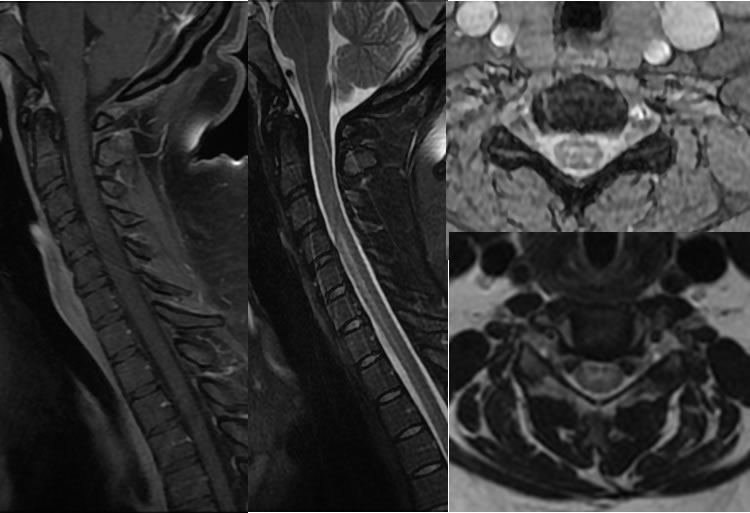
Multiple MRI images of the thoracic spine demonstrate long segment patchy ventral midline thoracic cord signal alteration without abnormal enhancement, syrinx, or cord expansion. The MRI images of the cervical spine demonstrate the superior extent of the cord signal abnormality which remains ventral and midline.
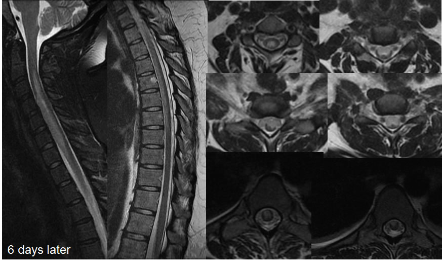
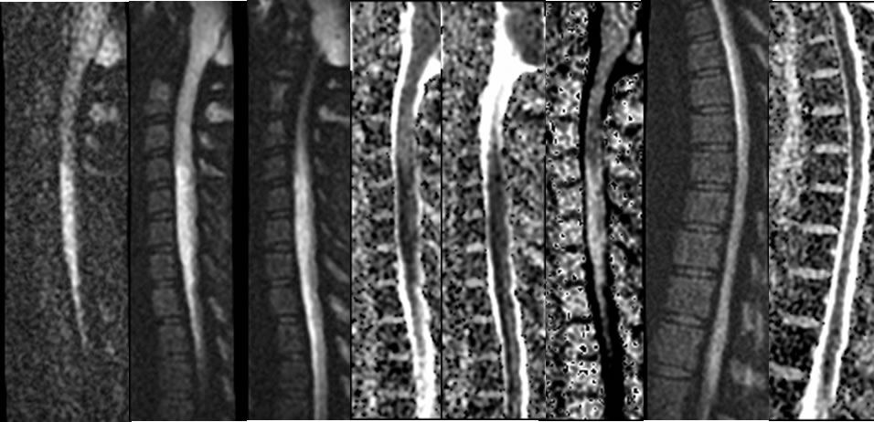
The follow up MRI images performed six days later demonstrate progressive cord signal alteration with a more wedge-shaped morphology on axial images. Mild cord expansion is now seen at lower cervical and upper thoracic levels. Diffusion weighted imaging of the spinal cord demonstrates abnormal restricted diffusion within the ventral midline spinal cord extending from mid cervical through midthoracic levels.
Discussion/Differential Diagnosis:
BACK TO
MAIN PAGE