EAC osteoma
Findings:
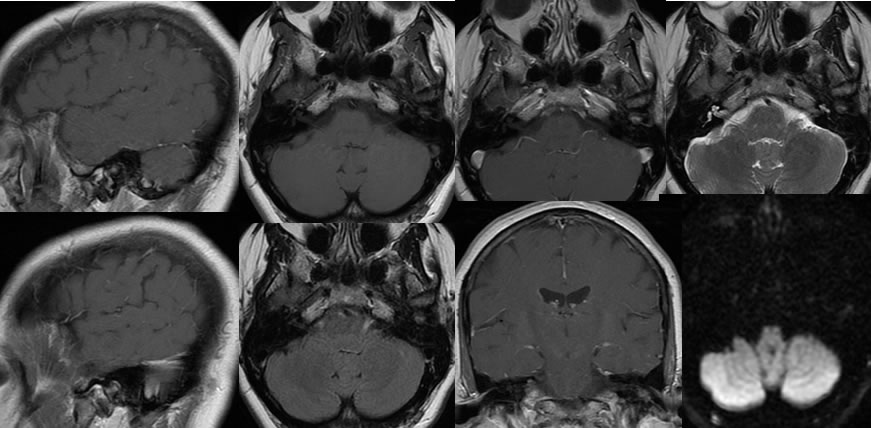
Multiple MRI images demonstrate a subtle nonspecific signal abnormality projected over the left external auditory canal. Mild enhancement and T2 hyperintensity is present. Diffusion characteristics are indeterminate.
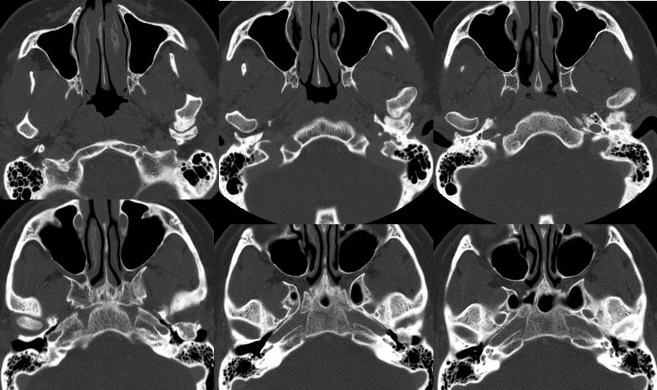
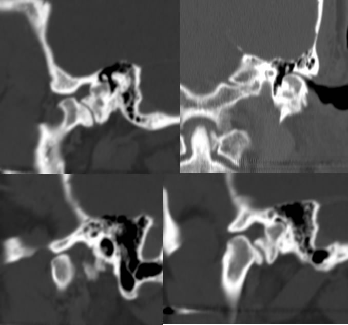
The CT images confirm that this is a long-standing ossified lesion with groundglass morphology internally, and adjacent bone remodeling including a lobulated projection from the left mandibular condyle which articulates with a portion of this lesion. The mass causes marked narrowing of the left external auditory canal.
Discussion/Differential Diagnosis:
BACK TO
MAIN PAGE