Benign Glomangioma with Ossification
Findings:
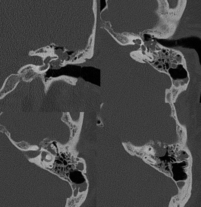
Temporal bone CT images demonstrate a multilobulated mass involving the left mesotympanum and epitympanum with mild erosive bone margins laterally and superomedially. A few small ossifications are seen within the mass. The mass opacifies the oval window niche and is inseparable from the facial nerve canal. There is associated inflammatory left mastoid air cell opacification with a fluid level.
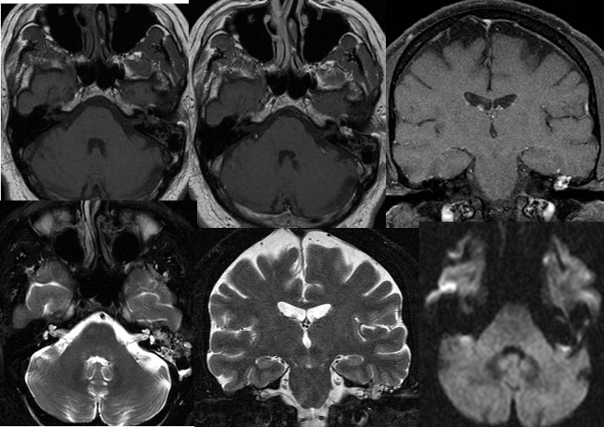
The MRI images confirm avid enhancement of the mass with relative T1 precontrast hyperintensity. Intermediate diffusion signal is seen in the region. The mass is well demarcated from inflammatory disease on the MRI images.
Discussion/Differential Diagnosis:
BACK TO
MAIN PAGE