Metastatic Lung Cancer
Findings:
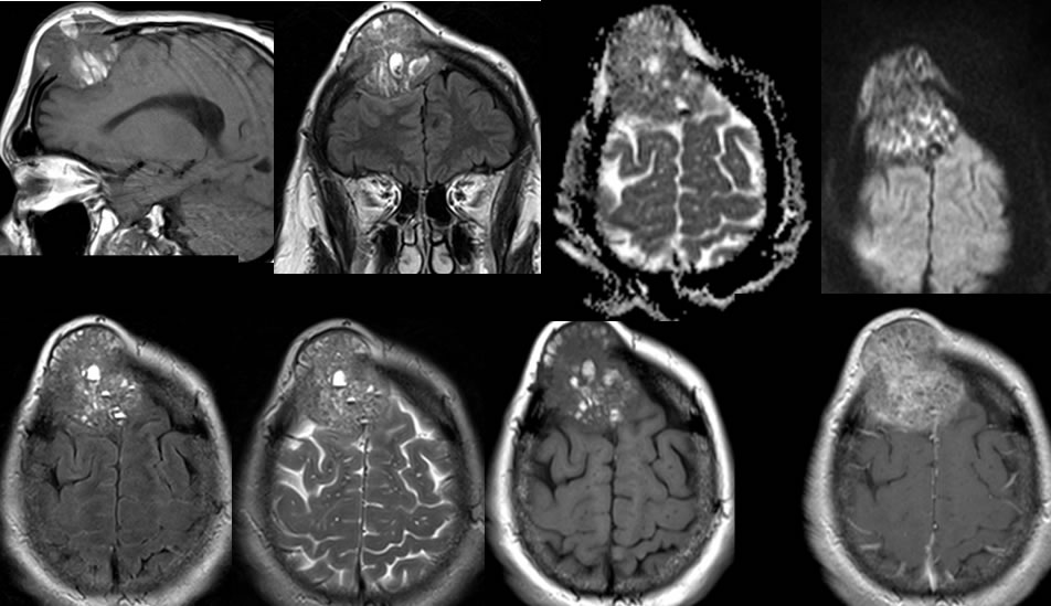
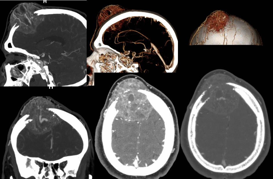
Multiple MRI and CT images including CTA reconstructions demonstrate a large destructive mass involving the right frontal calvarium. The mass has large extracranial and intracranial components but is overall well-defined and does not directly invade adjacent brain parenchyma or the subcutaneous fat of the scalp. The mass is hypervascular with multiple bizarre abnormal vessels within the lesion, multiple small cystic or centrally necrotic zones, as well as multiple fluid fluid levels seen on MRI. The mass has complex signal characteristics with mixed zones of T1 and T2 hyperintensity and hypointensity, with very heterogeneous enhancement.
Discussion/Differential Diagnosis:
BACK TO
MAIN PAGE