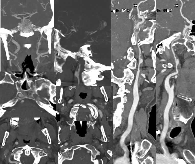
Grade 4 LICA Dissection
Findings:
Multiple CTA images demonstrate marked luminal irregularity and multifocal stenosis of the left internal carotid. A linear filling defect is seen at the C1-2 level on axial images. An irregular outpouching of contrast is present in this region due to a pseudoaneurysm. Distally at the level of the petrous cavernous ICA junction, there is a rounded occlusive filling defect. There is also subtle luminal irregularity of the right internal carotid with a small lateral outpouching seen on the coronal MIP image.
Discussion/Differential Diagnosis:
BACK TO
MAIN PAGE