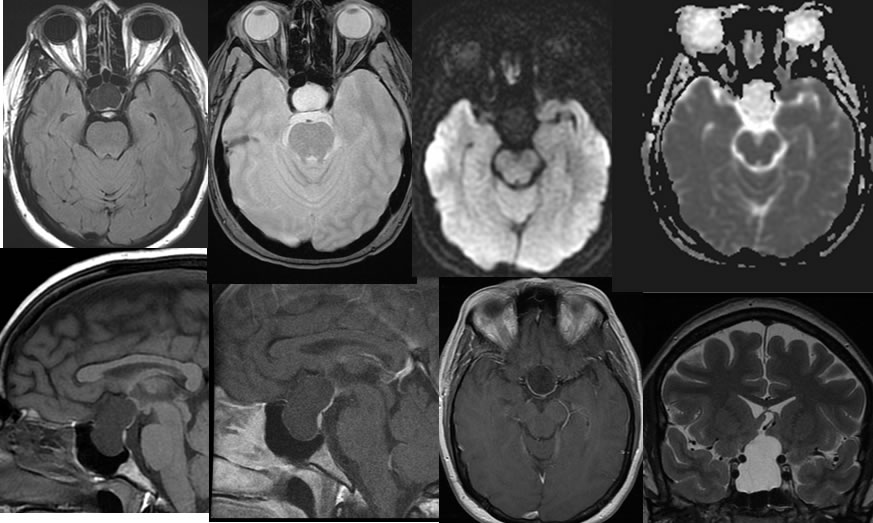
Rathke's Cleft Cyst
Findings:
Multiple MRI images demonstrate a large expansile fluid signal lesion expanding the sella and extending into the suprasellar cistern. The cystic lesion demonstrates signal nearly isointense to CSF. Uniform linear enhancing tissue is displaced peripherally along the posterior margin of the lesion, but there is no complete rim enhancement. The optic chiasm is not well identified, likely displaced and compressed by the lesion. There is no associated wall nodularity or cavernous sinus invasion. There is slight splaying of the internal carotids in this region without stenosis or encasement.
Discussion/Differential Diagnosis:
BACK TO
MAIN PAGE
