Squamous Cell Carcinoma, EX Inverting papilloma
Findings:
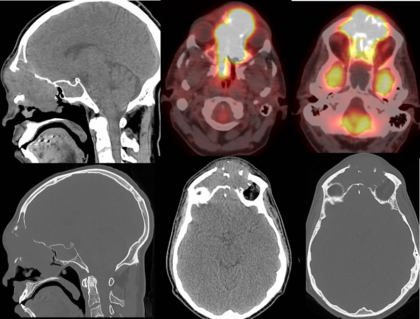
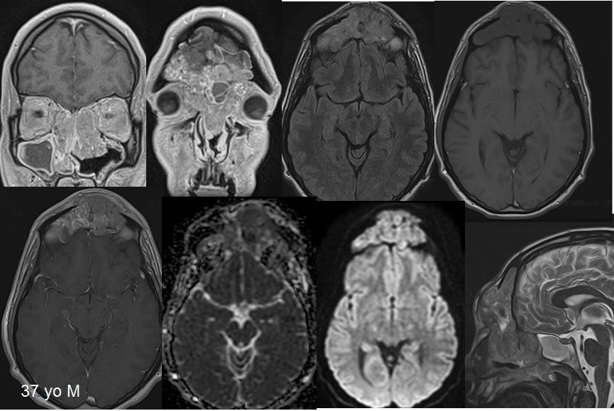
CT and PET CT images demonstrate a large destructive FDG avid soft tissue mass involving the frontal and ethmoid sinuses. There is extensive destruction of the frontal and ethmoid sinuses, including the posterior wall of the left frontal sinus. The MRI images demonstrate no direct parenchymal invasion or intracranial vasogenic edema. The mass demonstrates heterogeneous enhancement, diffusion restriction, and T2 hypointensity indicating a cellular lesion. There is superimposed postobstructive inflammatory disease in the right maxillary sinus with uniform peripheral enhancing mucosal thickening. There is also mild mucosal thickening in the left maxillary sinus. There is otherwise a relatively smooth expansile appearance of the bilateral frontal sinuses extending into the ethmoid regions. Superimposed mild bone remodeling is present indicating at least a component of a long-standing process. Postobstructive opacification of the sphenoid sinus is also identified. At the level of the destructive defect in the posterior wall of the left frontal sinus, there is adjacent dural invasion.
Discussion/Differential Diagnosis:
BACK TO
MAIN PAGE