CNS vasculitis
Findings:
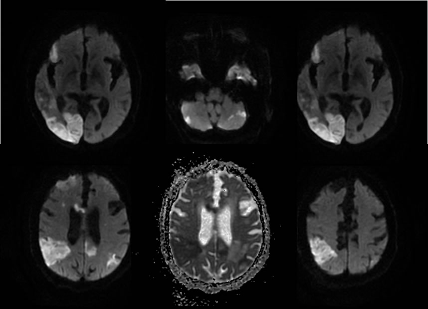
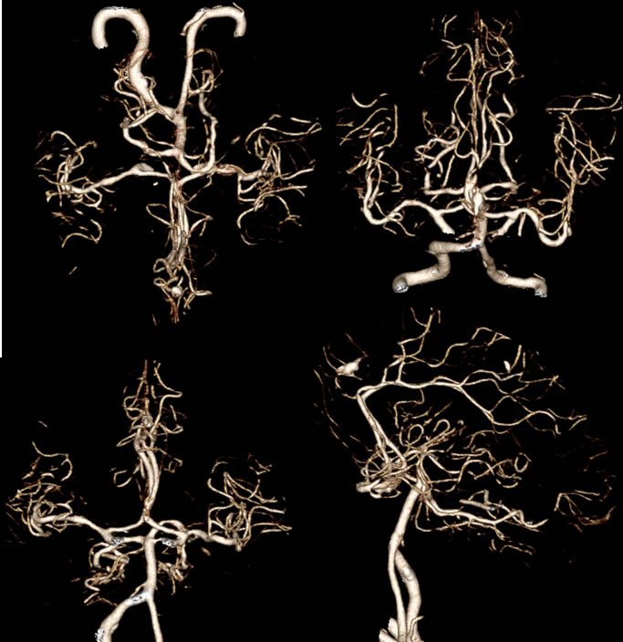
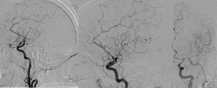
Diffusion weighted images demonstrate bilateral acute cerebral infarcts largest in the right parietooccipital region. This is superimposed on remote infarction in the left frontal lobe and confluent white matter disease. The CTA and DSA images demonstrate diffuse multifocal small vessel luminal irregularity with multifocal high-grade stenosis involving distal branches of the anterior, middle, and posterior cerebral arteries. Of note, no significant narrowing or luminal irregularity of proximal large vessels is seen, including the basilar artery, vertebral arteries, and the middle cerebral arteries. The proximal aspects of the anterior cerebral arteries also appear essentially normal.
Discussion/Differential Diagnosis:
BACK TO
MAIN PAGE