Type 2 Glomus AVM
Findings:
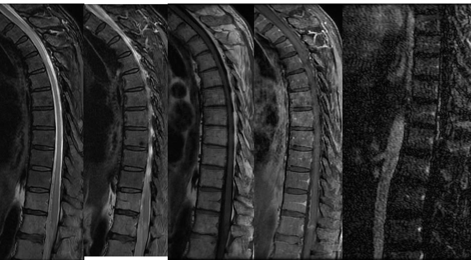
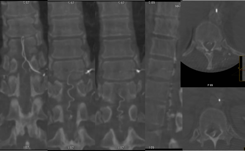
Sagittal MRI images of the thoracic spine demonstrate a nodular lesion at the level of the conus which demonstrates abnormal enhancement. Abnormal vascularity extends along the ventral margin of the spinal cord and there is mild surrounding edema in the spinal cord. A sagittal time resolved spinal arteriogram image demonstrates brisk AV shunting along the ventral margin of the spinal cord. A 3-D spinal conventional arteriogram with selective injection of the left T10 intercostal artery opacifies the artery of Adamceiwicz with the hairpin turn. There is arterial opacification of an intramedullary nidus at the level of the conus with tortuous early draining veins along the ventral margin of the spinal cord.
Discussion/Differential Diagnosis:
BACK TO
MAIN PAGE