Squamous Cell Carcinoma
Findings:
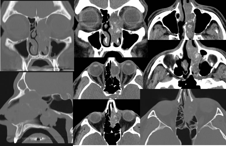
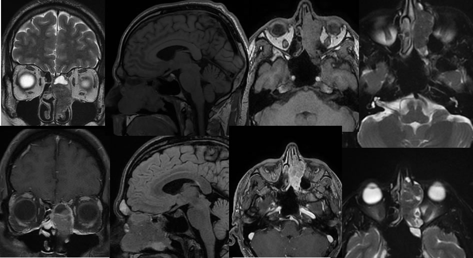
Multiple CT images demonstrate a heterogeneous mass within the left nasoethmoid region with associated relatively aggressive bone destruction and very little if any bone remodeling. The mass destroys the nasal septum, medial orbital wall, and ethmoid septae. Spotty calcifications are seen within the mass. Erosion of the planum sphenoidale is seen on the sagittal images. Multiple MRI images confirm the presence of a T2 hypointense enhancing mass within the left ethmoid region. Surrounding zones of postobstructive inflammatory disease are better demonstrated on MRI compared to CT, including postobstructive frontal sinus disease.
Discussion/Differential Diagnosis:
BACK TO
MAIN PAGE