Hemorrhagic Pituitary Adenoma
Findings:
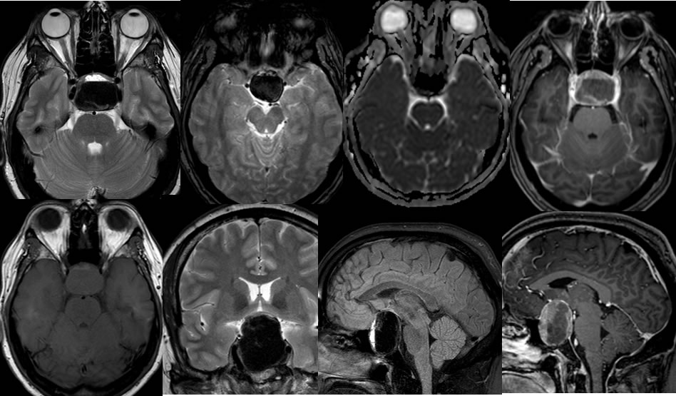
Multiple MRI images demonstrate a large complex cystic mass within the sella and suprasellar cistern, with significant expansion of the sella. The mass is well defined with significant superior displacement and compression of the optic chiasm, lateral splaying of the internal carotids, and no definite cavernous sinus invasion. The mass has a large hypointense fluid fluid level on the T2 weighted imaging, with heterogeneous signal on T1. Non-dependent T1 hyperintense fluid is present and there is mild irregular peripheral enhancement associated with the mass.
Discussion/Differential Diagnosis:
BACK TO
MAIN PAGE