Pituitary Adenoma, Cavernous Sinus Recurrence
Findings:
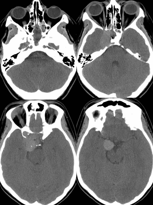
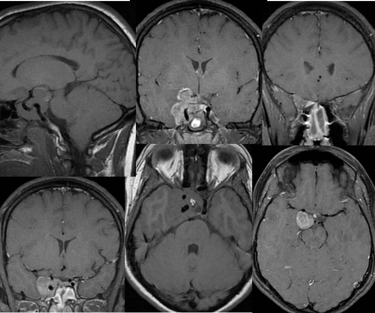
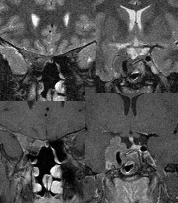
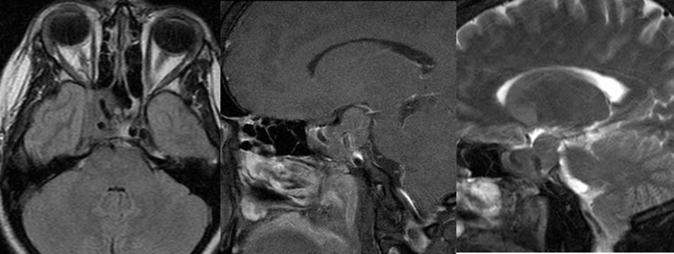
CT images demonstrate heterogeneous low-density opacification of the sphenoid sinus which is presumably related to previous transspheniodal surgery as confirmed on MRI. A hyperdense mass involves the right cavernous sinus on the CT, which demonstrates heterogeneous enhancement on MRI and encases the right internal carotid without significant narrowing. A lobulated zone of T1 precontrast hyperintensity on the axial image projected over the sella is most compatible with fat packing from the previous surgery. Inflammatory mucosal thickening within the sphenoid defect is incompletely included on the MRI. An additional linear zone of intense peripheral enhancement is associated with the right suprasellar component, which is inseparable from the hypothalamus and the medial temporal lobe. The mass also has subtle extent into the right orbital apex and demonstrates relative T2 hypointensity.
Discussion/Differential Diagnosis:
BACK TO
MAIN PAGE