Optic Nerve Sheath Meningioma
Findings:
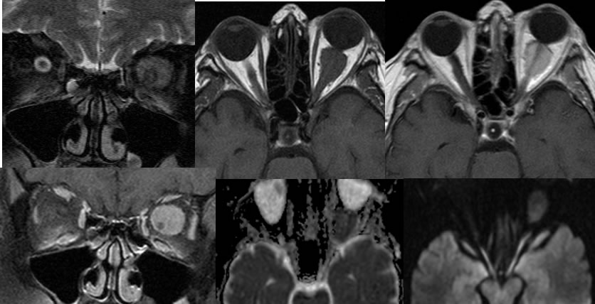
Multiple MRI images demonstrate a homogeneously enhancing mass within the intraconal left orbit with associated mild proptosis. The mass demonstrates mild restricted diffusion. A normal caliber optic nerve is seen encased by the lesion on the coronal images. Thickening of the optic nerve sheath is also visible on the coronal T2 weighted imaging.
Discussion/Differential Diagnosis:
BACK TO
MAIN PAGE