Pial AVM
Findings:
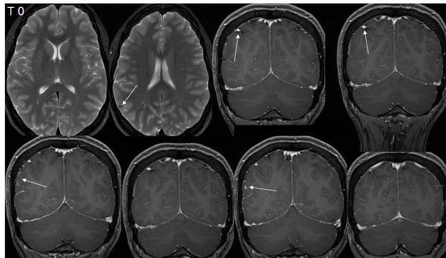
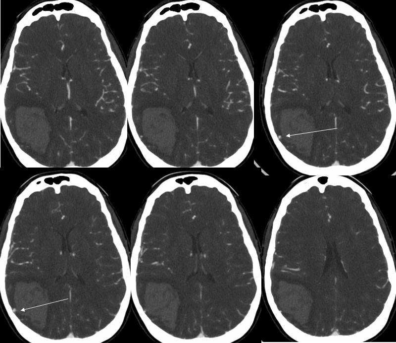
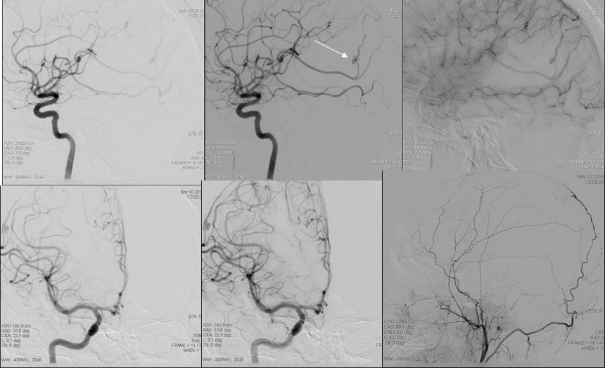
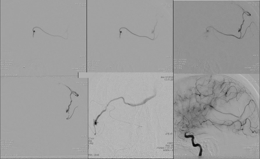
The baseline MRI images demonstrate a round well-defined hypointensity projected over the right parietal cortex shown by the arrow. Additional arrows demarcate an asymmetrically enlarged vein over the right parietal convexity. CTA images perform two years after the MRI demonstrate a large parenchymal hemorrhage within the right parietal lobe with a globular zone of enhancement laterally shown by the arrows at the level of the right parietal cortex. DSA images demonstrate a small nidus with AV shunting over the right parietal lobe, fed by a single right M4 branch and draining into a single cortical vein. The draining cortical vein demonstrates stenosis in the midportion. The final DSA image demonstrates occlusive embolization of the AVM.
Discussion/Differential Diagnosis:
BACK TO
MAIN PAGE