Squamous Cell Carcinoma, frontal sinus
Findings:
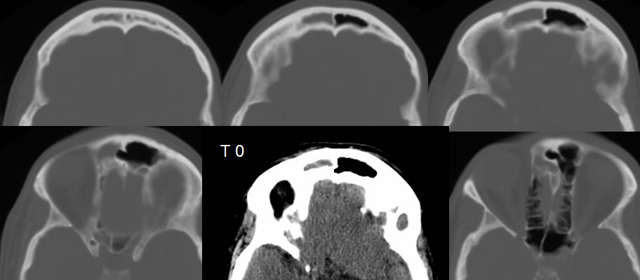
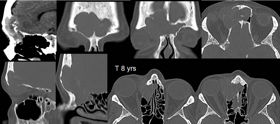
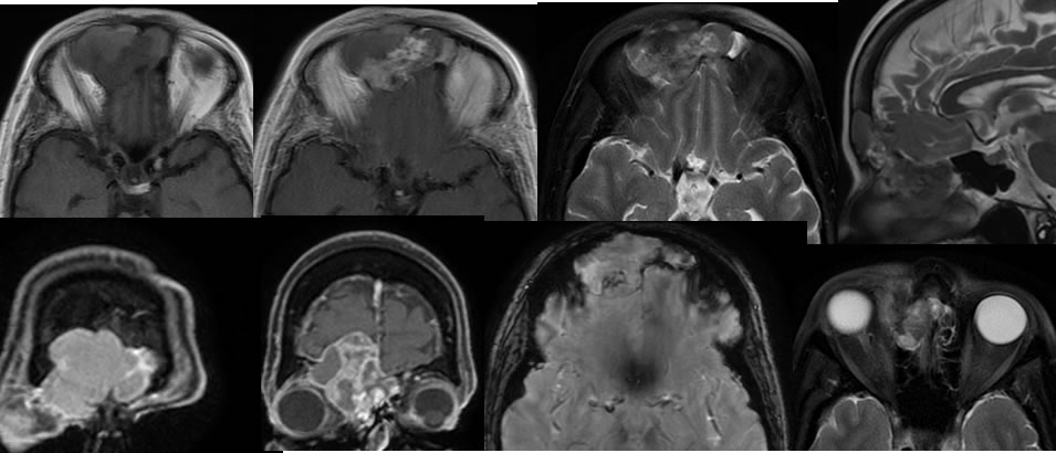
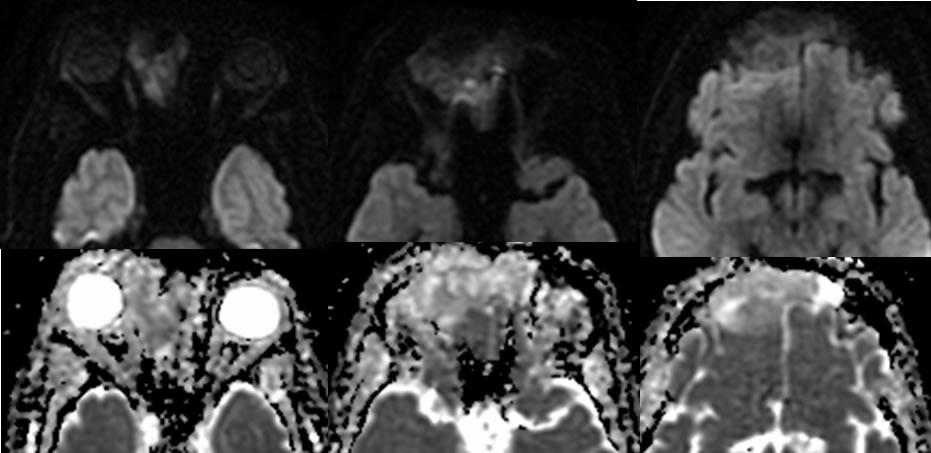
Baseline CT images demonstrate nonspecific complete opacification of the right frontal sinus with no definite expansile or destructive characteristics, most compatible chronic sinusitis. Minimal sclerosis and thickening of the frontal sinus walls is present. Images eight years later demonstrate development of a large destructive mass in this region which appears somewhat centered on the posterior wall of the right frontal sinus near the frontoethmoid junction. There is postobstructive opacification of the frontal sinus more superiorly with T1 hyperintense secretions that are decreased in T2 signal. The enhancing mass extends intracranially and involves the dura but does not directly invade the brain. No intracranial vasogenic edema is present. Intermediate diffusion characteristics are present, with some zones demonstrating true restricted diffusion along the posterior margin and other areas related to T2 shine through phenomenon.
Discussion/Differential Diagnosis:
BACK TO
MAIN PAGE