Myoepithelial Rich Pleomorphic Adenoma
Findings:
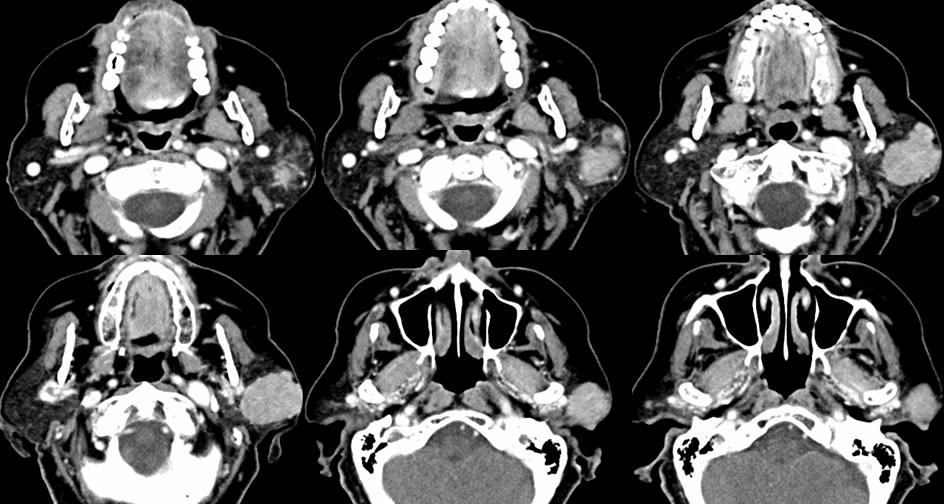
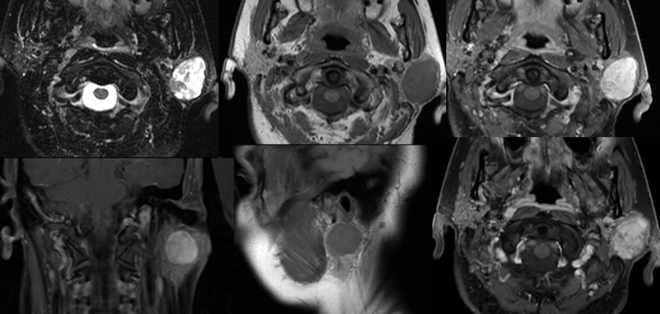
Contrast enhanced neck CT images demonstrate a relatively homogeneously enhancing slightly irregular multilobulated mass in the superficial lobe of the left parotid gland. The mass extends laterally to the level of the skin surface without definite disruption of the skin surface. The MRI images confirm intense enhancement of the mass, with heterogeneous T2 hyperintensity and slight hyperintensity to muscle on T1 weighted imaging.
Discussion/Differential Diagnosis:
BACK TO
MAIN PAGE