L5-S1 disc extrusion, resolves with conservative therapy
Findings:
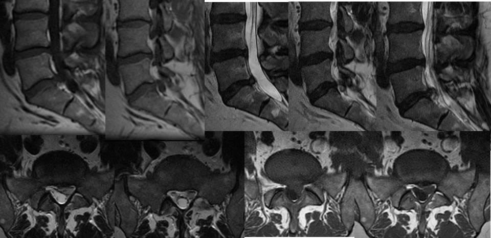
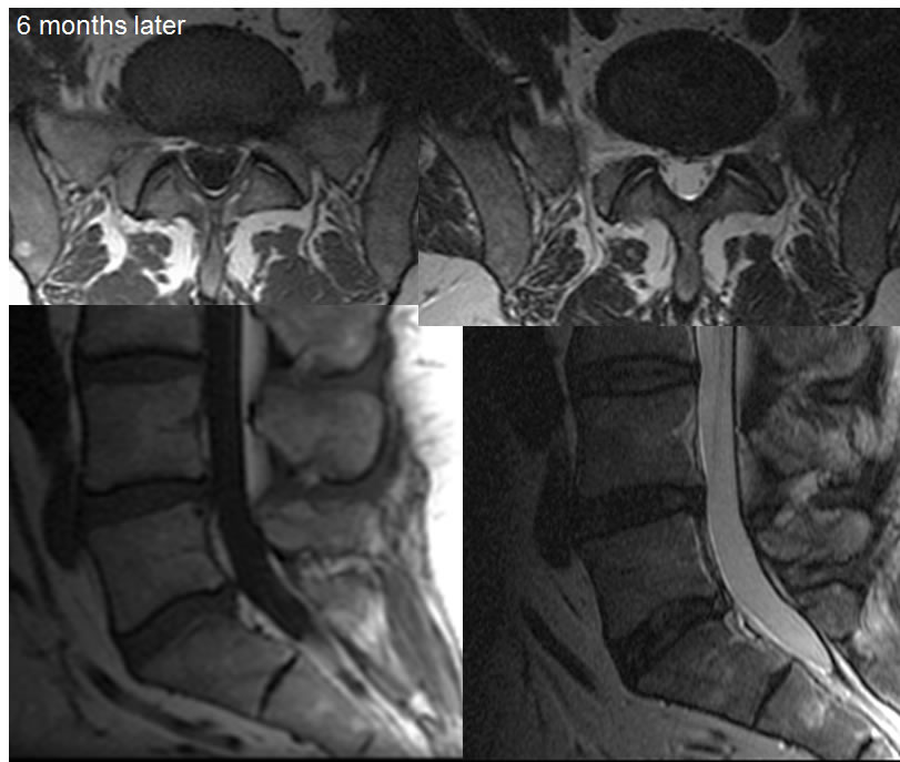
Multiple MRI images demonstrate a left central inferiorly extending disc extrusion at L5-S1 causing displacement of the left S1 nerve root and mild thecal sac compression. The extruded disc demonstrates relative T2 hyperintensity indicating that it may be recent. Six months later and without evidence of surgery, this disc extrusion has totally resolved with resolution of mass effect on the thecal sac. There is now a small noncompressive central disc protrusion and partial annular fissure at this level.
Discussion/Differential Diagnosis:
BACK TO
MAIN PAGE