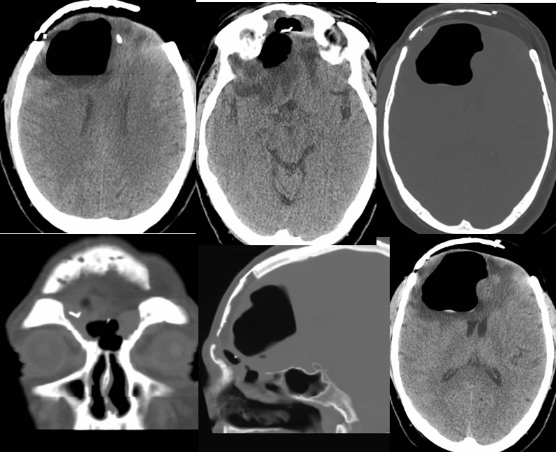
Surgical site dehiscence with pneumocephalus
Findings:
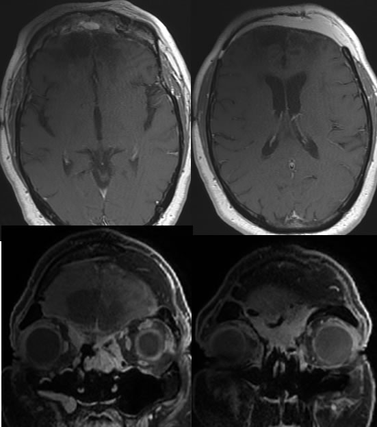
Baseline MRI images demonstrate postoperative findings of bifrontal craniectomy, reconstruction, and fat graft placement. Encephalomalacia and gliosis is present in the bilateral frontal lobes with volume loss. There is nonspecific enhancing tissue within the bilateral frontoethmoid regions and the right maxillary sinus, in part likely representing inflammatory disease. Mild reactive dural enhancement is present.
Follow-up CT images demonstrate development of a large gas collection in the right frontal lobe which on the coronal images is contiguous with the frontal sinus defect. A few tiny zones of dependent hyperdensity are present due to hemorrhage or other debris. The vasogenic edema is difficult to compare precisely but may be increased in the frontal lobes.
Discussion/Differential Diagnosis:
BACK TO
MAIN PAGE