Cholesteatoma
Findings:
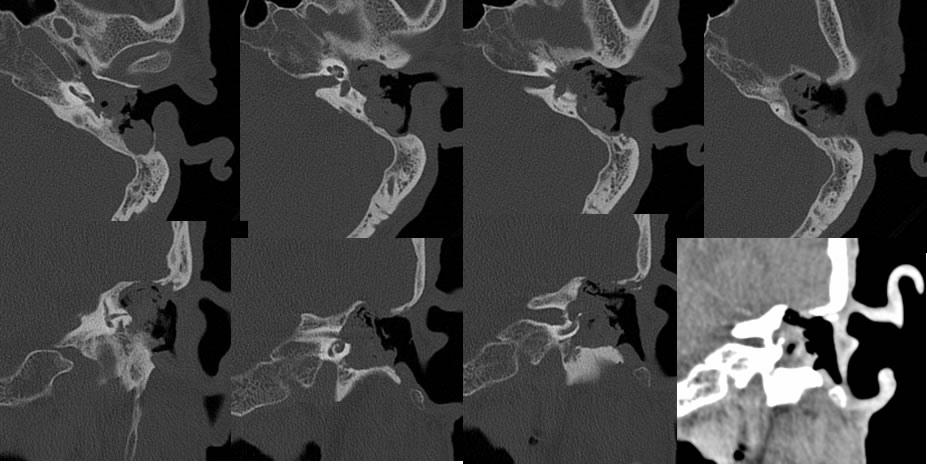
Multiple temporal bone CT images demonstrate a destructive but overall well-defined mass within the left extra auditory canal, middle ear cavity, and mastoid antrum. Large portions of the left otic capsule are destroyed, including a portion of the vestibule and the lateral semicircular canal. The included cochlea does not appear disrupted, but there is some marked thinning along the basal turn seen on coronal imaging. The soft tissue window demonstrates overall relative low density of this process. The lateral margin of the destructive lesion is very irregular with lobulated foci of debris and desquamed material.
Discussion/Differential Diagnosis:
BACK TO
MAIN PAGE