EAC Cholesteatoma and malignant otitis externa
Findings:
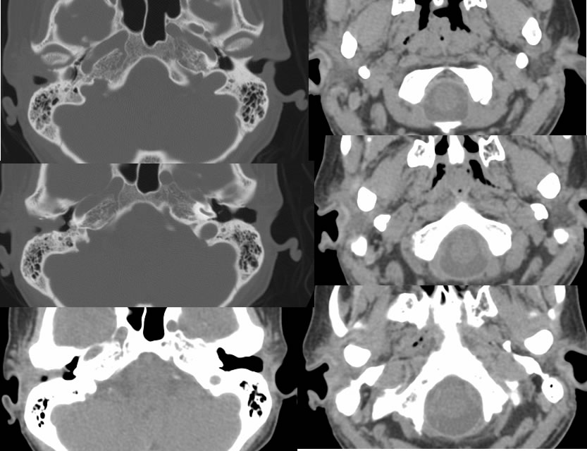
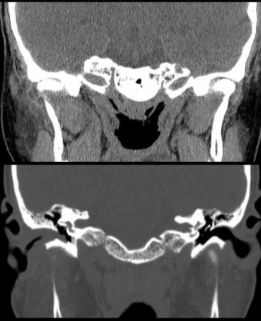
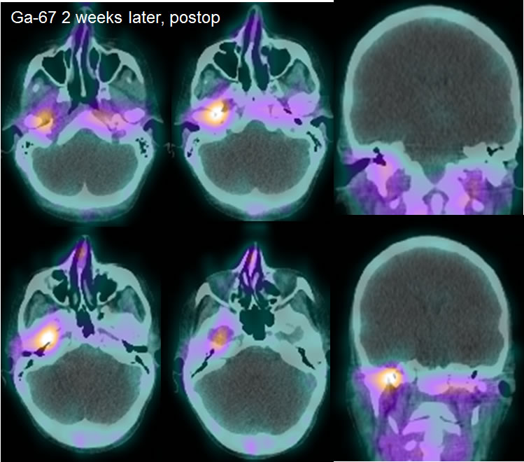
Multiple temporal bone CT images demonstrate complete opacification of the right external auditory canal with slightly irregular erosive margins. Mild to moderate opacification of right mastoid air cells present. Poorly defined hazy soft tissue opacity is present in the right peristyloid region and extending into the subcutaneous tissues, with obscured soft tissue planes adjacent to the right TM joint. Postoperative gallium scan images after resection of the external auditory canal mass demonstrate abnormal uptake within the right petrous bone and medial margin of the condylar fossa. No abnormal uptake is seen in the residual external auditory canal or adjacent soft tissues.
Discussion/Differential Diagnosis:
BACK TO
MAIN PAGE