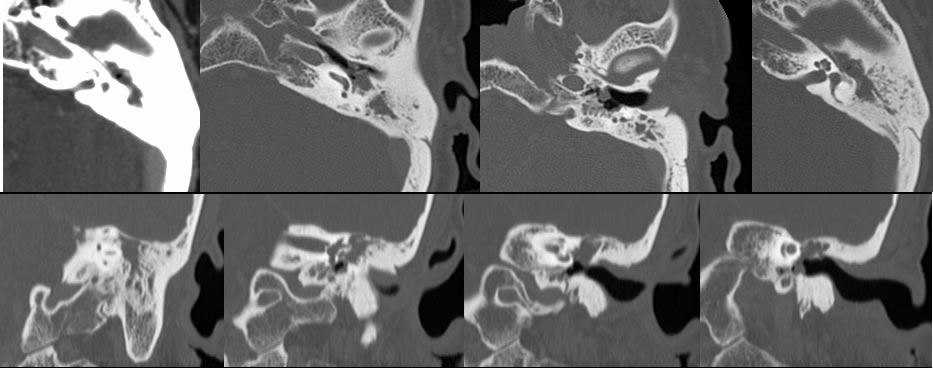
Cholesteatoma
Findings:
Multiple temporal bone CT images demonstrate opacification of the left mastoid air cells and middle ear cavity with medial retraction of the left tympanic membrane. No normal ossicles are visible. There is focal dehiscence in the lateral margin of the lateral semicircular canal. Additional lobulated soft tissue opacity extends more inferiorly along the retracted tympanic membrane. The scutum is slightly indistinct.
Discussion/Differential Diagnosis:
BACK TO
MAIN PAGE
