
Cholesteatoma
Findings:
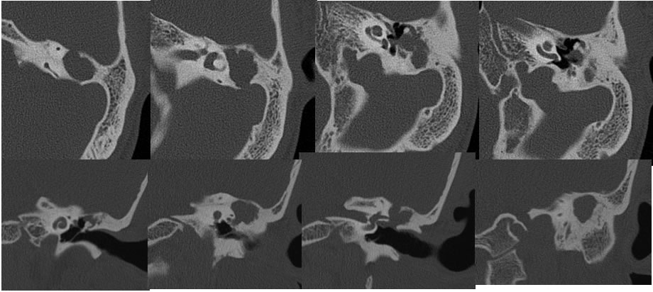
Multiple temporal bone CT images demonstrate expansile low density destructive opacification of the left mastoid antrum and epitympanum , associated with mild thickening of the tympanic membrane, and partial erosion of the incus. There is a focal defect in the lateral margin of the left lateral semicircular canal. There is additional dehiscence in the tegmen tympani and the sigmoid sinus bone plate. The included hypotympanum does not appear significantly opacified. Well defined scalloped destructive bone margins are present. The left mastoid air cells are otherwise underpneumatized.
Discussion/Differential Diagnosis:
BACK TO
MAIN PAGE