Benign Chondro-osseous Neoplasm
Findings:
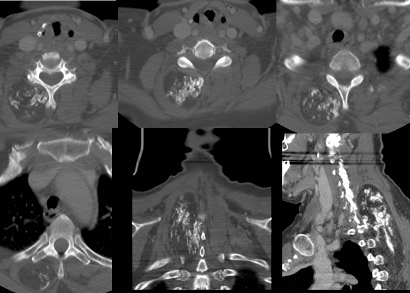
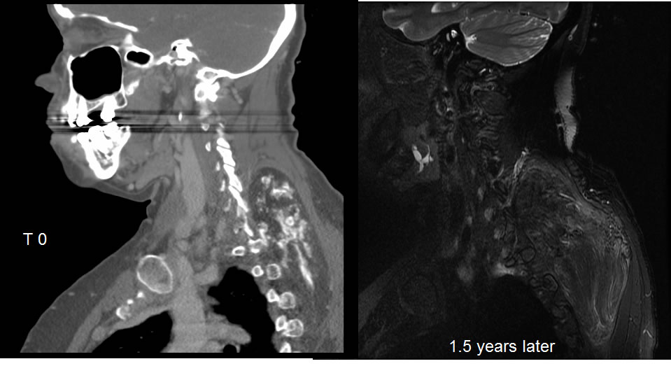
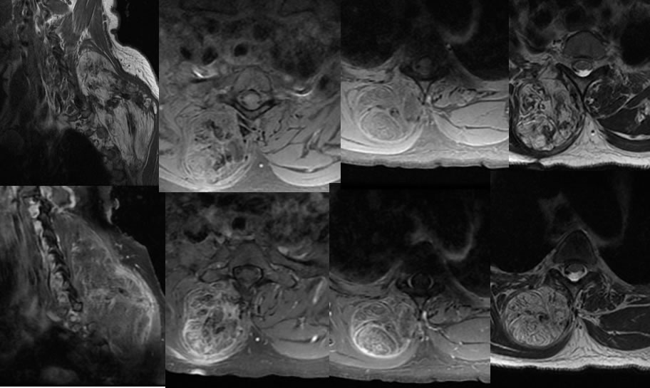
Multiple CT images with contrast demonstrate a large well-defined fat and calcium density mass located within the right paraspinal muscular structures extending from lower cervical through upper thoracic levels. No nodular zones of abnormal enhancement are seen on the CT images. The MRI images confirm linear and patchy zones of enhancement within the mass with no focal dominant nodularity. Zones of low signal on MRI correlate with the calcifications on CT. The mass has overall fat composition with zones of fat saturation. Some portions of the mass do not completely saturate with suppression of fat signal.
Discussion/Differential Diagnosis:
BACK TO
MAIN PAGE