Paraganglioma, recurrent
Findings:
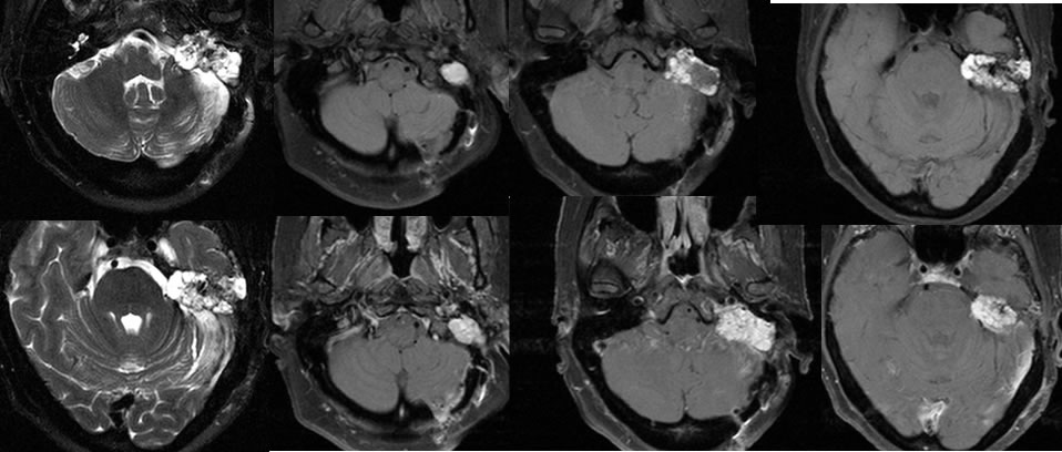
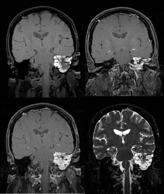
Multiple MRI images demonstrate postoperative findings of right retromastoid craniotomy. A multilobulated intensity enhancing mass with T2 hyperintensity involves the residual left petrous bone with intracranial extent. A zone of absent enhancement within the lateral margin of the mass could be a postoperative defect or findings of previous therapy with decreased enhancement. Numerous tiny T2 hypointensities within the mass could represent flow voids. There is a superimposed small amount of encephalomalacia and gliosis in the lateral margin of the left cerebellum with minimal reactive dural enhancement at the craniotomy site.
Discussion/Differential Diagnosis:
BACK TO
MAIN PAGE