Chondroblastic Osteosarcoma
Findings:
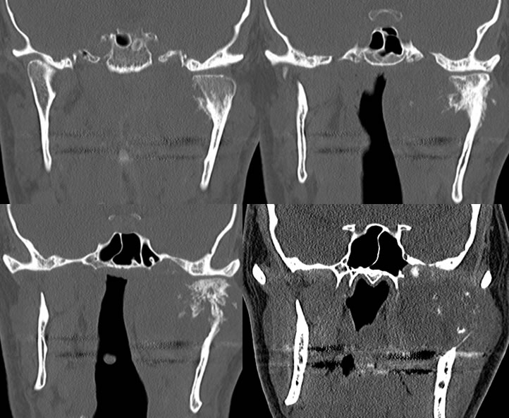
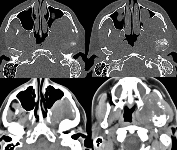
CT bone window images demonstrate remodeling of the left mandibular condyle associated with spiculated periosteal reaction in the subcondylar region. An associated overall low density infiltrative soft tissue mass is present extending into the masticator space with thinning and anterior bowing of the left maxillary sinus posterior wall. Post obstructive opacification of left mastoid air cells is present with thickening of soft tissues in the left nasopharynx on CT. The nasopharyngeal airway is slightly displaced to the right of midline. Superimposed mucous retention cysts are present in the paranasal sinuses.
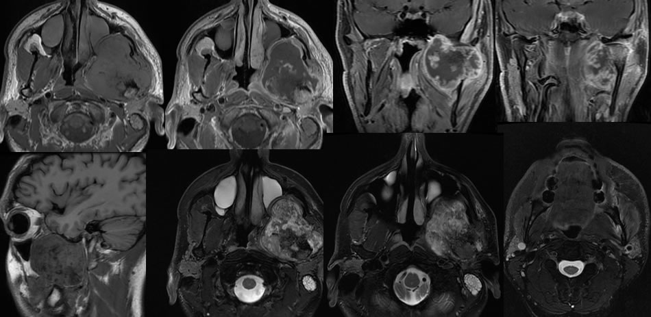
The MRI images demonstrate a very poorly defined perioherally enhancing and centrally necrotic soft tissue mass surrounding the right mandible with abnormal T2 hyperintensity within the left inferior alveolar canal. The mass has heterogeneous zones of low signal intensity in part correlating with the zones of periosteal reaction on CT. There is scalloped destruction of the left skull base on CT with dehiscence in the lateral margin of the left sphenoid sinus.
Discussion/Differential Diagnosis:
BACK TO
MAIN PAGE