EAC fibroosteoma
Findings:
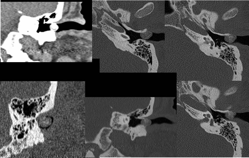
Multiple magnified CT Tempra bone images demonstrate a partially ossified well-defined lesion partially obstructing the left external auditory canal. Components of low density soft tissue and spotty calcifications are present in this region. There is no associated destruction of the external auditory canal.
Discussion/Differential Diagnosis:
BACK TO
MAIN PAGE