Recurrent Epidermoid
Findings:
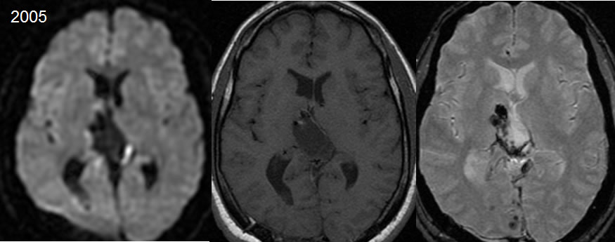
2005 postoperative MRI images demonstrate a surgical defect within the region of the cavum velum interpositum and posterior third ventricle. A linear focus of diffusion restriction is seen along the left lateral margin of the surgical defect. Spotty hemorrhagic changes are also seen along the surgical margins and along the right occipital parasagittal region.
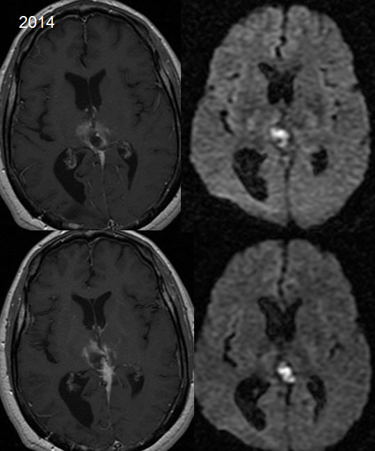
2014 images demonstrate contraction of the surgical defect with development of enhancement around the surgical cavity and a focus of diffusion restriction centrally.
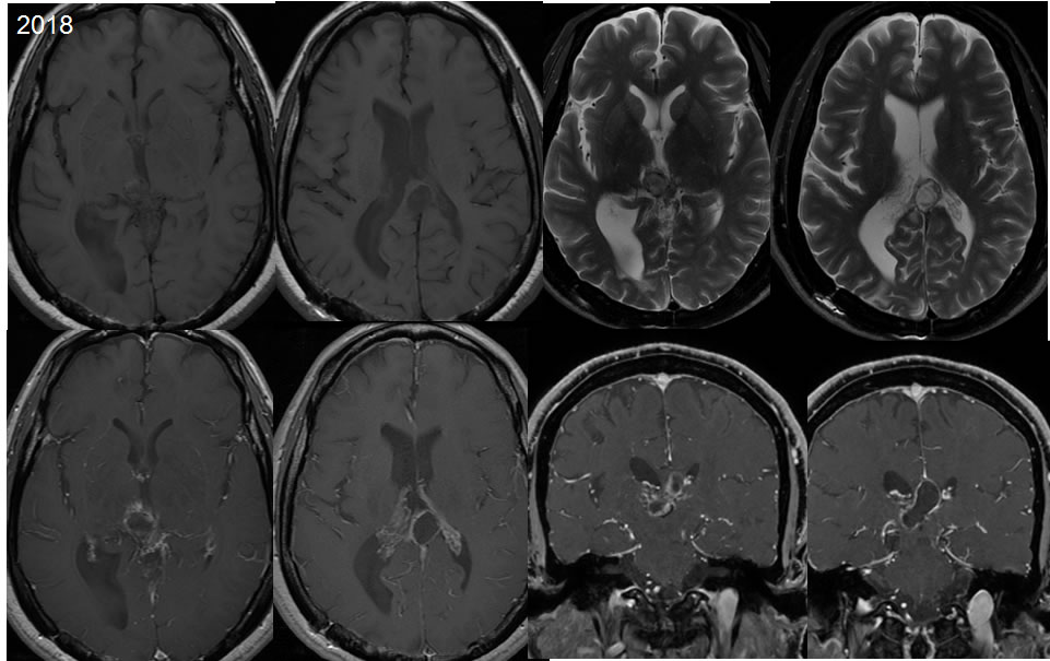
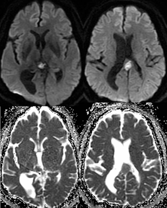
On the 2018 images, there is further subtle contraction of the surgical defect with diminished enhancement peripherally. There is a persistent zone of diffusion restriction within the surgical defect which is similar to slightly increased.
Discussion/Differential Diagnosis:
BACK TO
MAIN PAGE