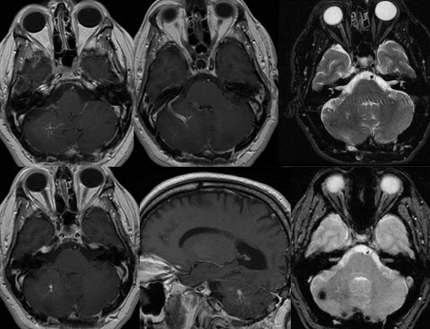
Developmental Venous Anomaly and Cavernoma
Findings:
Multiple MRI images demonstrate a large venous structure in the right cerebellum with branching tributaries medially due to a developmental venous anomaly. On the T2 weighted imaging, there is a superimposed focus of ringlike hypointensity laterally in the right cerebellum which demonstrates blooming on the gradient echo image. Postoperative findings of suboccipital craniotomy are also present for an abnormality not shown on these images.
Discussion/Differential Diagnosis:
BACK TO
MAIN PAGE