
PRES
Findings:
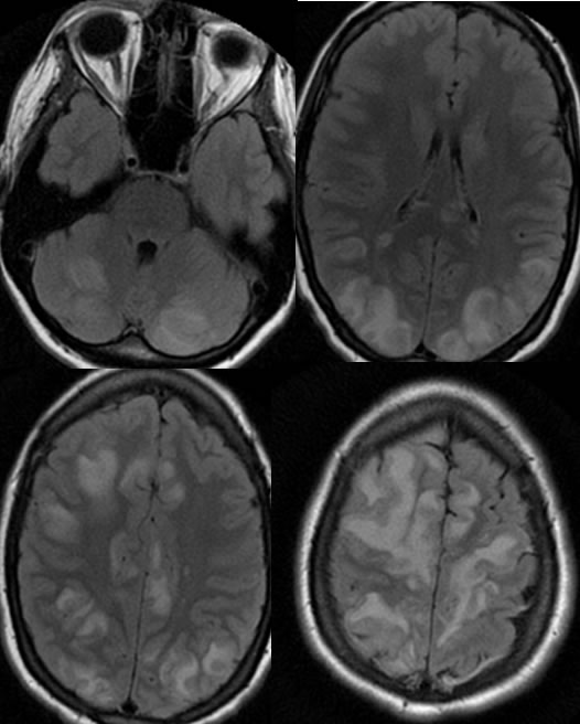
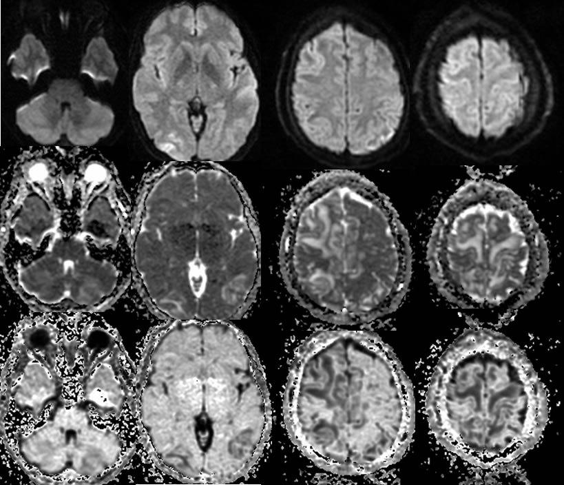
Axial FLAIR images demonstrate extensive bilateral frontal and parietooccipital cortical and subcortical signal abnormalities which also nearly symmetrically involve the cerebellar hemispheres. Mild sulcal effacement is present in the regions of signal alteration. The diffusion weighted images have a heterogeneous appearance with true restricted diffusion in the right occipital lobe and other areas of diffusion hypointensity in the subcortical white matter. ADC and exponential maps demonstrate facilitated diffusion in a patchy distribution throughout the cerebral white matter.
Discussion/Differential Diagnosis:
BACK TO
MAIN PAGE

