
PSP
Findings:
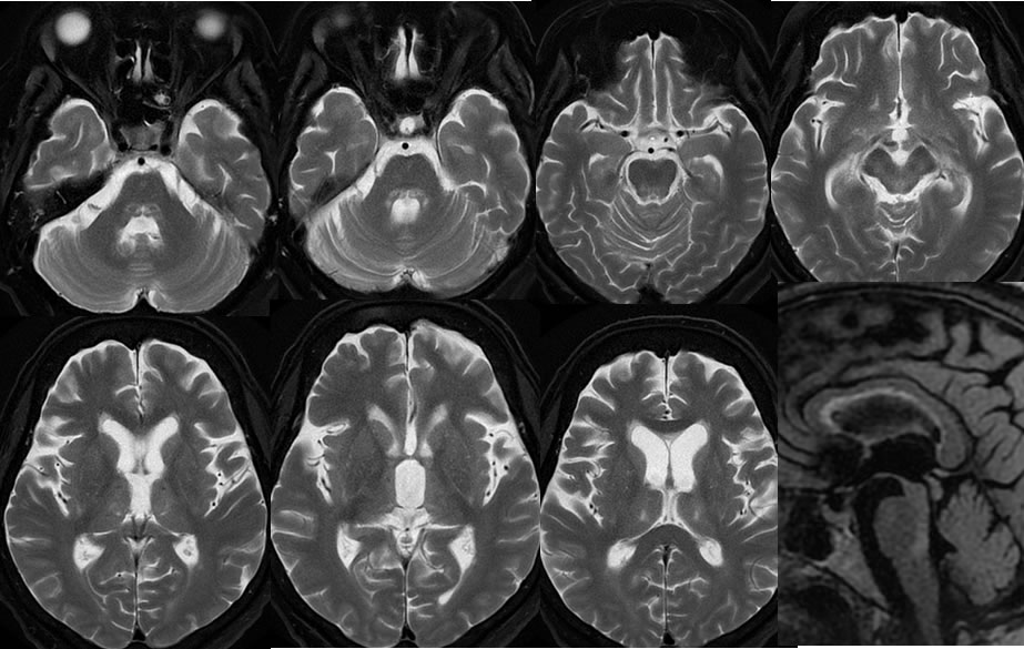
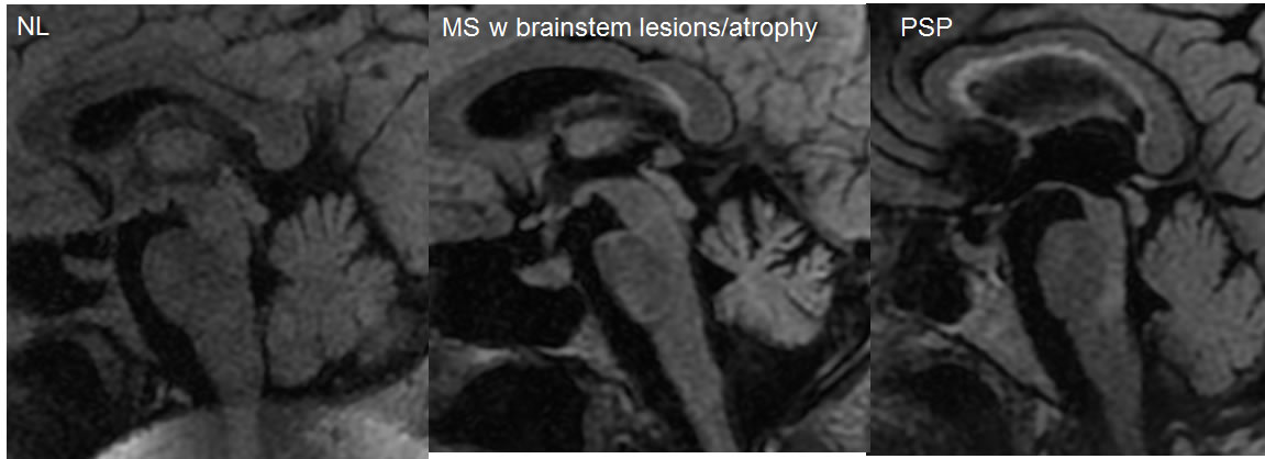
Multiple MRI images demonstrate selective atrophy of the midbrain with dilation of the third ventricle. The interpeduncular cistern is widened. Symmetric T2 hyperintensity is located in the dorsal pons and dorsal midbrain extending into the posterior thalami. The sagittal FLAIR image demonstrates more optimally the selective midbrain atrophy, with examples of normal midbrain volume for comparison.
Discussion/Differential Diagnosis:
BACK TO
MAIN PAGE

