Right Mesial Temporal Sclerosis
Findings:
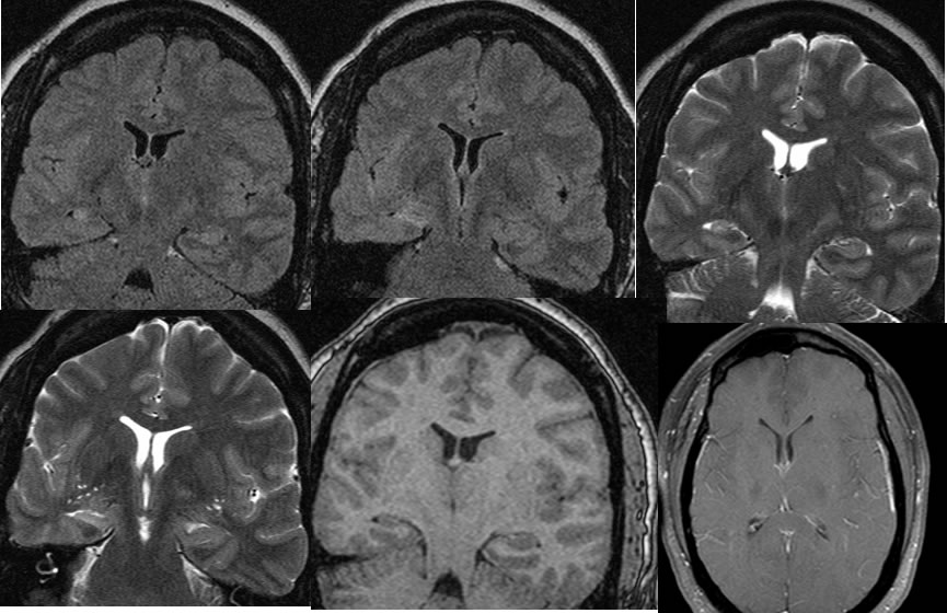
MRI seizure protocol images demonstrate asymmetric volume loss and T2 FLAIR hyperintensity within the right hippocampus. The difference in volume between the left hippocampus and the right is striking, and there is also a symmetric volume loss of the entire right cerebral hemisphere with hyperpneumatization of the right frontal sinus.
Discussion/Differential Diagnosis:
BACK TO
MAIN PAGE