PRES with hemorrhage and infarcts
Findings:
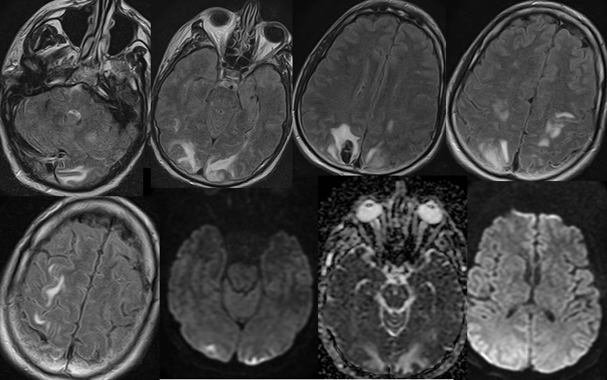
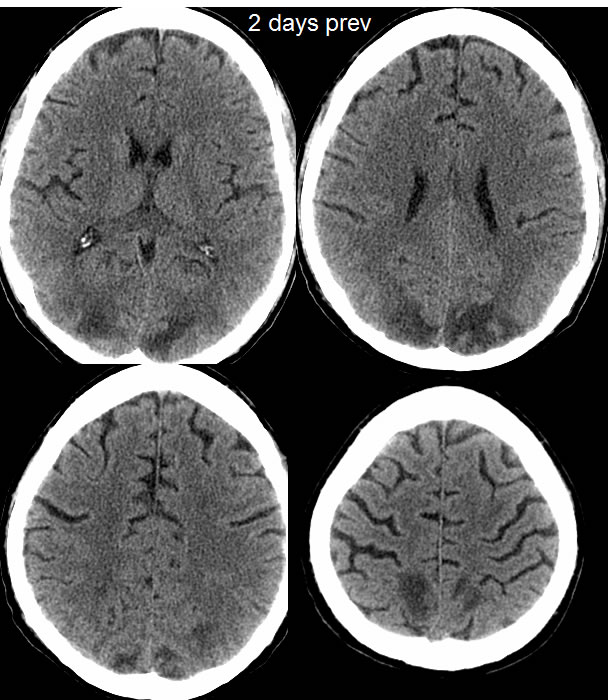
Multiple MRI images demonstrate patchy nearly symmetric FLAIR hyperintensities involving the bilateral parietooccipital regions, involving cortex and subcortical portions. Superimposed parenchymal hematoma is present in the right parietal parasagittal region.Restricted diffusion is present within the far peripheral occipital cortex bilaterally, with no other definite diffusion restriction. The signal abnormalities generally correlate with hypodensities on CT performed two days prior. Of note, the parenchymal hematoma is not visible previously. Mild sulcal effacement t is present in the region of signal abnormality. There is superimposed nonspecific white matter disease.
Discussion/Differential Diagnosis:
BACK TO
MAIN PAGE