

CSF hypotension
Findings:
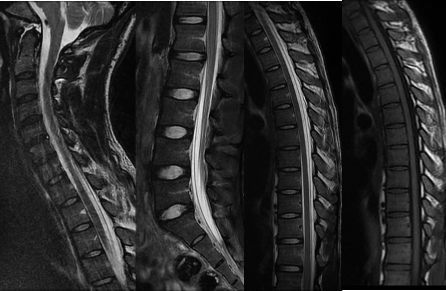
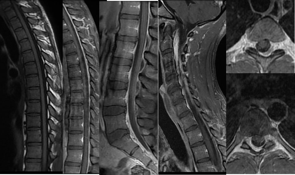
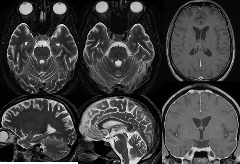
Sagittal MRI images of the spine demonstrate prominence of the dorsal epidural space at the thoracic levels and prominence of the ventral epidural space at lumbar levels with the appearance of extraaxial fluid collections. Following gadolinium administration, there is diffuse enhancement and congestion within the spinal epidural space with no significant mass effect on the thecal sac. The MRI images of the brain demonstrate absence of CSF space within the optic nerve sheaths with mild prominence of the extraaxial spaces. Mild diffuse dural enhancement is present after gadolinium administration. The dural sinuses have a rounded distended configuration.
Discussion/Differential Diagnosis:
BACK TO
MAIN PAGE


